 Explore
 Validate
 Learn
Learn Western blot
Antibody data
- Antibody Data
- Antigen structure
- References [1]
- Comments [0]
- Validations
- Western blot [2]
- Immunocytochemistry [1]
- Immunohistochemistry [4]
- Other assay [1]
Submit
Validation data
Reference
Comment
Report error
- Product number
- MA5-32160 - Provider product page

- Provider
- Invitrogen Antibodies
- Product name
- PRMT5 Recombinant Rabbit Monoclonal Antibody (ST51-06)
- Antibody type
- Monoclonal
- Antigen
- Synthetic peptide
- Reactivity
- Human, Mouse, Rat
- Host
- Rabbit
- Isotype
- IgG
- Antibody clone number
- ST51-06
- Vial size
- 100 µL
- Concentration
- 1 mg/mL
- Storage
- Store at 4°C short term. For long term storage, store at -20°C, avoiding freeze/thaw cycles.
Submitted references Protein Arginine Methyltransferase 5 as a Therapeutic Target for KRAS Mutated Colorectal Cancer.
Shifteh D, Sapir T, Pahmer M, Haimowitz A, Goel S, Maitra R
Cancers 2020 Jul 28;12(8)
Cancers 2020 Jul 28;12(8)
No comments: Submit comment
Supportive validation
- Submitted by
- Invitrogen Antibodies (provider)
- Main image

- Experimental details
- Western blot analysis of PRMT5 in A431 cell lysate using a PRMT5 Monoclonal antibody (Product # MA5-32160) at a dilution of 1:1,000.

- Submitted by
- Invitrogen Antibodies (provider)
- Main image

- Experimental details
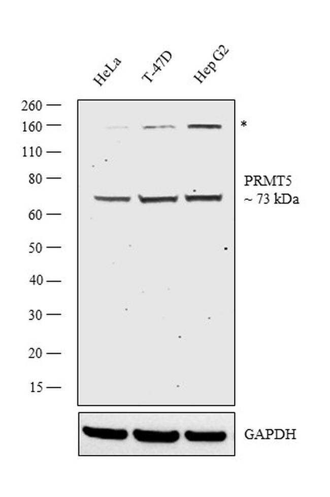
- Western blot analysis was performed on nuclear enriched cell extract (30 µg lysate) of HeLa (Lane 1), T-47D (Lane 2), and Hep G2 (Lane 3). The blot was probed with Anti-PRMT5 Monoclonal Antibody (Product # MA5-32160, 1:1000 dilution) and detected by chemiluminescence using Goat anti-Rabbit IgG (H+L) Superclonal™ Secondary Antibody, HRP conjugate (Product # A27036, 0.25 µg/mL, 1:4000 dilution). A 73 kDa band corresponding to PRMT5 was observed in all cell lines tested with a *non-specific band at ~160 kDa.
Supportive validation
- Submitted by
- Invitrogen Antibodies (provider)
- Main image

- Experimental details
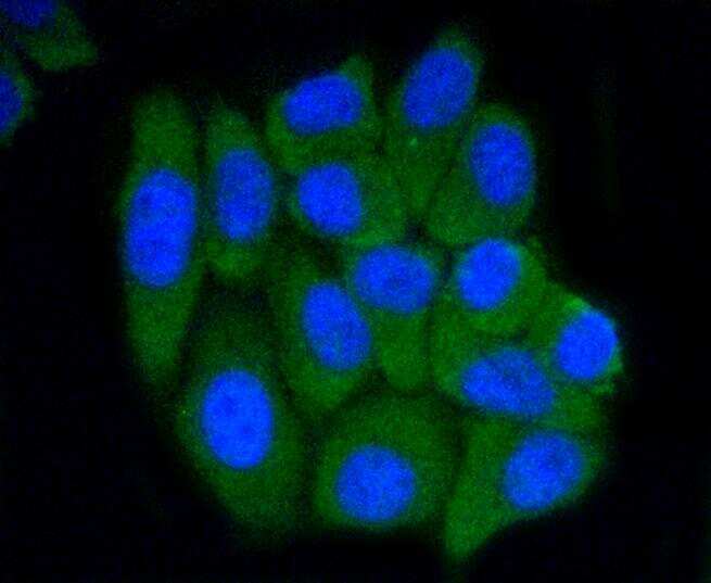
- Immunocytochemical analysis of PRMT5 in HepG2 cells using a PRMT5 Monoclonal antibody (Product # MA5-32160) as seen in green. The nuclear counter stain is DAPI (blue). Cells were fixed in paraformaldehyde, permeabilised with 0.25% Triton X100/PBS.
Supportive validation
- Submitted by
- Invitrogen Antibodies (provider)
- Main image
- Experimental details
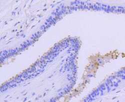
- Immunohistochemical analysis of PRMT5 of paraffin-embedded Human breast carcinoma tissue using a PRMT5 Monoclonal antibody (Product #MA5-32160). Counter stained with hematoxylin.
- Submitted by
- Invitrogen Antibodies (provider)
- Main image
- Experimental details
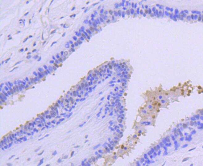
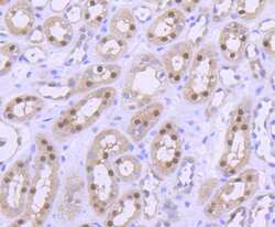
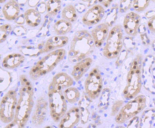
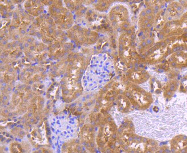
- Immunohistochemical analysis of PRMT5 of paraffin-embedded Human kidney tissue using a PRMT5 Monoclonal antibody (Product #MA5-32160). Counter stained with hematoxylin.
- Submitted by
- Invitrogen Antibodies (provider)
- Main image

- Experimental details
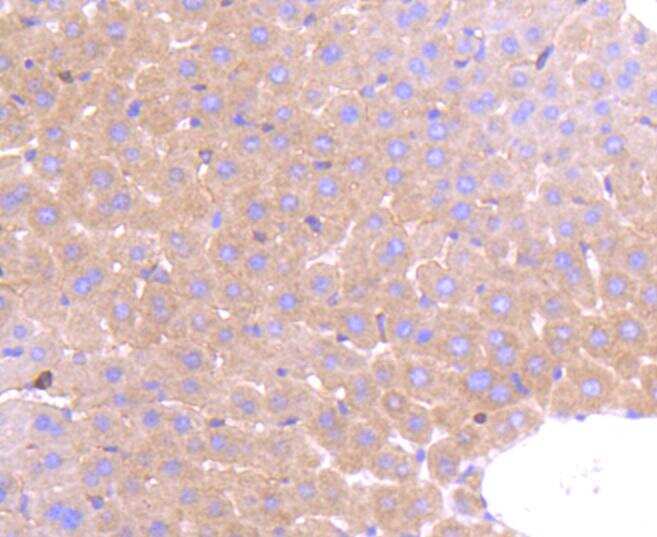
- Immunohistochemical analysis of PRMT5 of paraffin-embedded Mouse liver tissue using a PRMT5 Monoclonal antibody (Product #MA5-32160). Counter stained with hematoxylin.
- Submitted by
- Invitrogen Antibodies (provider)
- Main image
- Experimental details
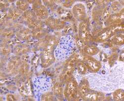
- Immunohistochemical analysis of PRMT5 of paraffin-embedded Mouse kidney tissue using a PRMT5 Monoclonal antibody (Product #MA5-32160). Counter stained with hematoxylin.
Supportive validation
- Submitted by
- Invitrogen Antibodies (provider)
- Main image

- Experimental details
- Figure 3 PRMT5 is shown to be further overexpressed in the KRAS mutant CRC cells at the translational level by Western blot analysis. ( a ) Western blot assay results displaying PRMT5 and corresponding beta-actin bands; ( b ) Quantified Western blot assay results showing that PRMT5 protein is 4.8-Fold ( p < 0.01) further overexpressed in the KRAS mutant CRC cells when compared to the KRAS WT CRC cells. Data are expressed as means + SD from two independent experiments using four CRC cell lines (2 KRAS mutant, 2 KRAS WT), as well as one normal colon cell line. The uncropped Western blot membranes can be seen in Figure S1 . Data for the individual cell lines can be seen in Figures S2 and S3 . * represents p < 0.05; ** represents p < 0.01.
