 Explore
 Validate
 Learn
Learn Western blot
 Immunocytochemistry
Antibody data
- Antibody Data
- Antigen structure
- References [3]
- Comments [0]
- Validations
- Western blot [1]
- Flow cytometry [1]
Submit
Validation data
Reference
Comment
Report error
- Product number
- MAB6166 - Provider product page

- Provider
- Novus Biologicals
- Product name
- Mouse Monoclonal EOMES Antibody
- Antibody type
- Monoclonal
- Description
- Protein A or G purified from hybridoma culture supernatant. Detects human EOMES in direct ELISAs. In direct ELISAs, no cross-reactivity with recombinant human (rh) Brachyury, rhEOMES (aa 1-115), recombinant mouse EOMES (aa 1-126), rhTBX2, 3, 5, 6, 18, or 20 is observed.
- Reactivity
- Human
- Host
- Mouse
- Isotype
- IgG
- Vial size
- 100 ug
- Concentration
- LYOPH
- Storage
- Use a manual defrost freezer and avoid repeated freeze-thaw cycles. 12 months from date of receipt, -20 to -70 degreesC as supplied. 1 month, 2 to 8 degreesC under sterile conditions after reconstitution. 6 months, -20 to -70 degreesC under sterile conditions after reconstitution.
Submitted references Reliability of human cortical organoid generation.
WNT signaling memory is required for ACTIVIN to function as a morphogen in human gastruloids.
Elf5-centered transcription factor hub controls trophoblast stem cell self-renewal and differentiation through stoichiometry-sensitive shifts in target gene networks.
Yoon SJ, Elahi LS, Pașca AM, Marton RM, Gordon A, Revah O, Miura Y, Walczak EM, Holdgate GM, Fan HC, Huguenard JR, Geschwind DH, Pașca SP
Nature methods 2019 Jan;16(1):75-78
Nature methods 2019 Jan;16(1):75-78
WNT signaling memory is required for ACTIVIN to function as a morphogen in human gastruloids.
Yoney A, Etoc F, Ruzo A, Carroll T, Metzger JJ, Martyn I, Li S, Kirst C, Siggia ED, Brivanlou AH
eLife 2018 Oct 12;7
eLife 2018 Oct 12;7
Elf5-centered transcription factor hub controls trophoblast stem cell self-renewal and differentiation through stoichiometry-sensitive shifts in target gene networks.
Latos PA, Sienerth AR, Murray A, Senner CE, Muto M, Ikawa M, Oxley D, Burge S, Cox BJ, Hemberger M
Genes & development 2015 Dec 1;29(23):2435-48
Genes & development 2015 Dec 1;29(23):2435-48
No comments: Submit comment
Supportive validation
- Submitted by
- Novus Biologicals (provider)
- Main image
- Experimental details
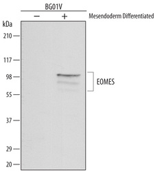
- Detection of Human EOMES by Western Blot. Western blot shows lysates of BG01V human embryonic stem cells untreated (-) or mesendoderm differentiated (+). PVDF membrane was probed with 1 µg/mL of Mouse Anti-Human EOMES Monoclonal Antibody (Catalog # MAB6166) followed by HRP-conjugated Anti-Mouse IgG Secondary Antibody (Catalog # HAF007). Specific bands were detected for EOMES at approximately 100 and 80 kDa (as indicated). This experiment was conducted under reducing conditions and using Immunoblot Buffer Group 1.

Supportive validation
- Submitted by
- Novus Biologicals (provider)
- Main image
- Experimental details
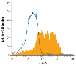
- Detection of EOMES in Differentiated BG01VHuman Cells by Flow Cytometry. BG01V human embryonic stem cells differentiated to mesendoderm were stained with Mouse Anti-Human EOMES Monoclonal Antibody (Catalog # MAB6166, filled histogram) or isotype control antibody (Catalog # MAB0041, open histogram), followed by Allo-phycocyanin-conjugated Anti-Mouse IgG Secondary Antibody (Catalog # F0101B). To facilitate intracellular staining, cells were fixed with paraformaldehyde and permeabilized with saponin.
