Explore
 Validate
 Learn
Learn Western blot
Antibody data
- Antibody Data
- Antigen structure
- References [1]
- Comments [0]
- Validations
- Western blot [1]
- Immunocytochemistry [1]
- Flow cytometry [1]
Submit
Validation data
Reference
Comment
Report error
- Product number
- AF6166 - Provider product page

- Provider
- R&D Systems
- Product name
- Human EOMES Antibody
- Antibody type
- Polyclonal
- Description
- Immunogen affinity purified. Detects human EOMES in Western blots.
- Reactivity
- Human
- Host
- Sheep
- Conjugate
- Unconjugated
- Antigen sequence
O95936- Isotype
- IgG
- Vial size
- 100 ug
- Concentration
- LYOPH
- Storage
- Use a manual defrost freezer and avoid repeated freeze-thaw cycles. 12 months from date of receipt, -20 to -70 °C as supplied. 1 month, 2 to 8 °C under sterile conditions after reconstitution. 6 months, -20 to -70 °C under sterile conditions after reconstitution.
Submitted references Reprogramming to pluripotency does not require transition through a primitive streak-like state.
Raab S, Klingenstein M, Möller A, Illing A, Tosic J, Breunig M, Kuales G, Linta L, Seufferlein T, Arnold SJ, Kleger A, Liebau S
Scientific reports 2017 Nov 29;7(1):16543
Scientific reports 2017 Nov 29;7(1):16543
No comments: Submit comment
Supportive validation
- Submitted by
- R&D Systems (provider)
- Main image
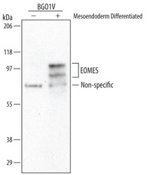
- Experimental details
- Detection of Human EOMES by Western Blot. Western blot shows lysates of BG01V human embryonic stem cells untreated (-) or mesoendoderm differentiated (+). PVDF Membrane was probed with 1 µg/mL of Human EOMES Antigen Affinity-purified Polyclonal Antibody (Catalog # AF6166) followed by HRP-conjugated Anti-Sheep IgG Secondary Antibody (Catalog # HAF016). Specific bands were detected for EOMES at approximately 85-105 kDa (as indicated). This experiment was conducted under reducing conditions and using Immunoblot Buffer Group 1.
Supportive validation
- Submitted by
- R&D Systems (provider)
- Main image

- Experimental details
- EOMES in mesoderm lineage cells differentiated from BG01V. EOMES was detected in immersion fixed BG01V human embryonic stem cells differentiated into mesoderm using Human EOMES Antigen Affinity-purified Polyclonal Antibody (Catalog # AF6166) at 10 µg/mL for 3 hours at room temperature. Cells were stained using the NorthernLights™ 557-conjugated Anti-Sheep IgG Secondary Antibody (red, upper panel; Catalog # NL010) and counterstained with DAPI (blue, lower panel). Specific staining was localized to nuclei. View our protocol for Fluorescent ICC Staining of Cells on Coverslips.

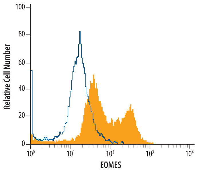
Supportive validation
- Submitted by
- R&D Systems (provider)
- Main image

- Experimental details
- Detection of EOMES in Differ-entiated BG01V Human Cells by Flow Cytometry. BG01V human embryonic stem cells differentiated to mesendoderm were stained with Sheep Anti-Human EOMES Antigen Affinity-purified Polyclonal Antibody (Catalog # AF6166, filled histogram) or isotype control antibody (Catalog # 5-001-A, open histogram), followed by Allophycocyanin-conjugated Anti-Sheep IgG Secondary Antibody (Catalog # F0127). To facilitate intracellular staining, cells were fixed with paraformaldehyde and permeabilized with saponin.