Explore
 Validate
 Learn
Learn Western blot
Antibody data
- Antibody Data
- Antigen structure
- References [2]
- Comments [0]
- Validations
- Western blot [1]
- Immunocytochemistry [1]
- Flow cytometry [1]
Submit
Validation data
Reference
Comment
Report error
- Product number
- MAB6166 - Provider product page

- Provider
- R&D Systems
- Product name
- Human EOMES Antibody
- Antibody type
- Monoclonal
- Description
- Protein A or G purified from hybridoma culture supernatant. Detects human EOMES in direct ELISAs. In direct ELISAs, no cross-reactivity with recombinant human (rh) Brachyury, rhEOMES (aa 1-115), recombinant mouse EOMES (aa 1-126), rhTBX2, 3, 5, 6, 18, or 20 is observed.
- Reactivity
- Human
- Host
- Mouse
- Conjugate
- Unconjugated
- Antigen sequence
O95936- Isotype
- IgG
- Antibody clone number
- 644730
- Vial size
- 100 ug
- Concentration
- LYOPH
- Storage
- Use a manual defrost freezer and avoid repeated freeze-thaw cycles. 12 months from date of receipt, -20 to -70 °C as supplied. 1 month, 2 to 8 °C under sterile conditions after reconstitution. 6 months, -20 to -70 °C under sterile conditions after reconstitution.
Submitted references WNT signaling memory is required for ACTIVIN to function as a morphogen in human gastruloids.
Elf5-centered transcription factor hub controls trophoblast stem cell self-renewal and differentiation through stoichiometry-sensitive shifts in target gene networks.
Yoney A, Etoc F, Ruzo A, Carroll T, Metzger JJ, Martyn I, Li S, Kirst C, Siggia ED, Brivanlou AH
eLife 2018 Oct 12;7
eLife 2018 Oct 12;7
Elf5-centered transcription factor hub controls trophoblast stem cell self-renewal and differentiation through stoichiometry-sensitive shifts in target gene networks.
Latos PA, Sienerth AR, Murray A, Senner CE, Muto M, Ikawa M, Oxley D, Burge S, Cox BJ, Hemberger M
Genes & development 2015 Dec 1;29(23):2435-48
Genes & development 2015 Dec 1;29(23):2435-48
No comments: Submit comment
Supportive validation
- Submitted by
- R&D Systems (provider)
- Main image
- Experimental details
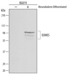
- Detection of Human EOMES by Western Blot. Western blot shows lysates of BG01V human embryonic stem cells untreated (-) or mesendoderm differentiated (+). PVDF membrane was probed with 1 µg/mL of Mouse Anti-Human EOMES Monoclonal Antibody (Catalog # MAB6166) followed by HRP-conjugated Anti-Mouse IgG Secondary Antibody (Catalog # HAF007). Specific bands were detected for EOMES at approximately 100 and 80 kDa (as indicated). This experiment was conducted under reducing conditions and using Immunoblot Buffer Group 1.

Supportive validation
- Submitted by
- R&D Systems (provider)
- Main image
- Experimental details
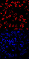
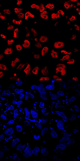
- EOMES in Mesodermally Differentiated BG01V Human Stem Cells. EOMES was detected in immersion fixed BG01V human embryonic stem cells differentiated into mesoderm using Human EOMES Monoclonal Antibody (Catalog # MAB6166) at 10 µg/mL for 3 hours at room temperature. Cells were stained using the NorthernLights™ 557-conjugated Anti-Mouse IgG Secondary Antibody (red, upper panel; Catalog # NL007) and counterstained with DAPI (blue, lower panel). Specific staining was localized to nuclei. View our protocol for Fluorescent ICC Staining of Cells on Coverslips.
Supportive validation
- Submitted by
- R&D Systems (provider)
- Main image

- Experimental details
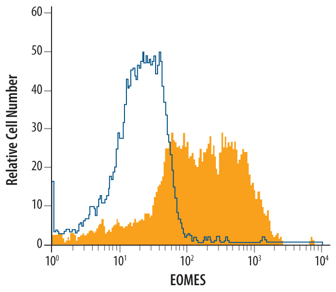
- Detection of EOMES in Differentiated BG01VHuman Cells by Flow Cytometry. BG01V human embryonic stem cells differentiated to mesendoderm were stained with Mouse Anti-Human EOMES Monoclonal Antibody (Catalog # MAB6166, filled histogram) or isotype control antibody (Catalog # MAB0041, open histogram), followed by Allo-phycocyanin-conjugated Anti-Mouse IgG Secondary Antibody (Catalog # F0101B). To facilitate intracellular staining, cells were fixed with paraformaldehyde and permeabilized with saponin.