 Explore
 Validate
 Learn
Learn Western blot
Antibody data
- Antibody Data
- Antigen structure
- References [5]
- Comments [0]
- Validations
- Western blot [2]
- Immunocytochemistry [1]
Submit
Validation data
Reference
Comment
Report error
- Product number
- MA5-15163 - Provider product page

- Provider
- Invitrogen Antibodies
- Product name
- Phospho-MYL9 (Ser19) Monoclonal Antibody (R.179.1)
- Antibody type
- Monoclonal
- Antigen
- Synthetic peptide
- Description
- It is not recommended to aliquot this antibody.
- Antibody clone number
- R.179.1
- Concentration
- 445 µg/mL
Submitted references Behind Brain Metastases Formation: Cellular and Molecular Alterations and Blood-Brain Barrier Disruption.
ADF and cofilin-1 collaborate to promote cortical actin flow and the leader bleb-based migration of confined cells.
Switching between blebbing and lamellipodia depends on the degree of non-muscle myosin II activity.
Doxorubicin induces detrusor smooth muscle impairments through myosin dysregulation, leading to a risk of lower urinary tract dysfunction.
Septin-dependent remodeling of cortical microtubule drives cell reshaping during epithelial wound healing.
Godinho-Pereira J, Garcia AR, Figueira I, Malhó R, Brito MA
International journal of molecular sciences 2021 Jun 30;22(13)
International journal of molecular sciences 2021 Jun 30;22(13)
ADF and cofilin-1 collaborate to promote cortical actin flow and the leader bleb-based migration of confined cells.
Ullo MF, Logue JS
eLife 2021 Jun 25;10
eLife 2021 Jun 25;10
Switching between blebbing and lamellipodia depends on the degree of non-muscle myosin II activity.
Ghosh I, Singh RK, Mishra M, Kapoor S, Jana SS
Journal of cell science 2021 Jan 13;134(1)
Journal of cell science 2021 Jan 13;134(1)
Doxorubicin induces detrusor smooth muscle impairments through myosin dysregulation, leading to a risk of lower urinary tract dysfunction.
Iguchi N, Dönmez Mİ, Carrasco A Jr, Wilcox DT, Pineda RH, Malykhina AP, Cost NG
American journal of physiology. Renal physiology 2019 Jul 1;317(1):F197-F206
American journal of physiology. Renal physiology 2019 Jul 1;317(1):F197-F206
Septin-dependent remodeling of cortical microtubule drives cell reshaping during epithelial wound healing.
Shindo A, Audrey A, Takagishi M, Takahashi M, Wallingford JB, Kinoshita M
Journal of cell science 2018 Jun 28;131(12)
Journal of cell science 2018 Jun 28;131(12)
No comments: Submit comment
Supportive validation
- Submitted by
- Invitrogen Antibodies (provider)
- Main image

- Experimental details
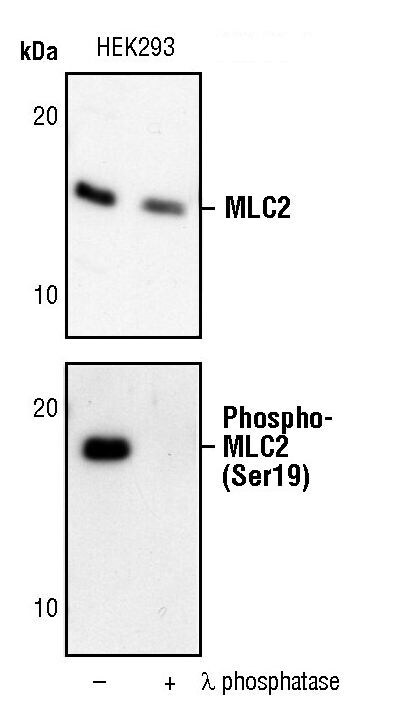
- Western blot analysis of Phospho-Myosin Light Chain 2 pSer19 in extracts from HEK293 cells, untreated (-) or lambda phosphatase-treated (+), using Phospho-Myosin Light Chain 2 pSer19 monoclonal antibody (Product # MA5-15163) (upper) or Phospho-Myosin Light Chain 2 (Ser19) monoclonal antibody (lower).
- Submitted by
- Invitrogen Antibodies (provider)
- Main image

- Experimental details
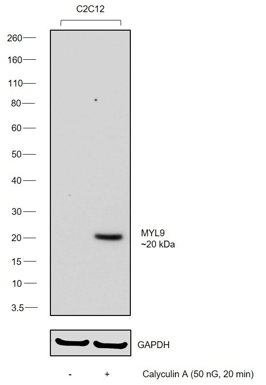
- Western blot was performed using Anti-Phospho-MYL9 (Ser19) Monoclonal Antibody (R.179.1) (Product # MA5-15163) and a ~20 kDa band corresponding to Myosin regulatory light polypeptide 9 was observed in C2C12 after treatment with Calyculin A. Whole cell extracts (40 µg lysate) of C2C12 (Lane 1) and Calyculin A treated C2C12 (Lane 2) were electrophoresed using NuPAGE™ 4-12% Bis-Tris Protein Gel (Product # NP0321BOX). Resolved proteins were then transferred onto a nitrocellulose membrane (Product # IB23001) by iBlot® 2 Dry Blotting System (Product # IB21001). The blot was probed with the primary antibody (1:1000 dilution) and detected by chemiluminescence with Goat anti-Mouse IgG (H+L) Superclonal™ Recombinant Secondary Antibody, HRP (Product # A28177,1:20000 dilution) using the iBright FL 1000 (Product # A32752). Chemiluminescent detection was performed using SuperSignal™ West Dura Extended Duration Substrate (Product # 34076).
Supportive validation
- Submitted by
- Invitrogen Antibodies (provider)
- Main image
- Experimental details
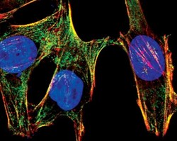
- Immunofluorescent analysis of Phospho-Myosin Light Chain 2 pSer19 in HeLa cells using a Phospho-Myosin Light Chain 2 pSer19 onoclonal antibody (Product # MA5-15163) (green). Actin filaments are labeled with a fluorescent red phalloidin. DNA is labeled using a fluorescent blue dye.
