 Explore
 Validate
 Learn
Learn Western blot
Antibody data
- Antibody Data
- Antigen structure
- References [1]
- Comments [0]
- Validations
- Western blot [1]
- Immunocytochemistry [2]
- Flow cytometry [1]
Submit
Validation data
Reference
Comment
Report error
- Product number
- MAB7106 - Provider product page

- Provider
- R&D Systems
- Product name
- Human ER beta/NR3A2 Antibody
- Antibody type
- Monoclonal
- Description
- Protein A or G purified from hybridoma culture supernatant. Detects human ER beta/NR3A2 in direct ELISAs and Western blot. In direct ELISA and Western blot, no cross-reactivity with recombinant human ER alpha is observed.
- Reactivity
- Human
- Host
- Mouse
- Conjugate
- Unconjugated
- Antigen sequence
Q92731- Isotype
- IgG
- Antibody clone number
- 733930
- Vial size
- 100 ug
- Concentration
- LYOPH
- Storage
- Use a manual defrost freezer and avoid repeated freeze-thaw cycles. 12 months from date of receipt, -20 to -70 °C as supplied. 1 month, 2 to 8 °C under sterile conditions after reconstitution. 6 months, -20 to -70 °C under sterile conditions after reconstitution.
Submitted references Estradiol-mediated improvements in adipose tissue insulin sensitivity are related to the balance of adipose tissue estrogen receptor α and β in postmenopausal women.
Park YM, Pereira RI, Erickson CB, Swibas TA, Cox-York KA, Van Pelt RE
PloS one 2017;12(5):e0176446
PloS one 2017;12(5):e0176446
No comments: Submit comment
Supportive validation
- Submitted by
- R&D Systems (provider)
- Main image
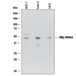
- Experimental details
- Detection of Human ER beta/NR3A2 by Western Blot. Western blot shows lysates of MCF-7 human breast cancer cell line, Saos-2 human osteosarcoma cell line, and T47D human breast cancer cell line. PVDF membrane was probed with 2 µg/mL of Mouse Anti-Human ER beta/NR3A2 Monoclonal Antibody (Catalog # MAB7106) followed by HRP-conjugated Anti-Mouse IgG Secondary Antibody (Catalog # HAF007). A specific band was detected for ER beta/NR3A2 at approximately 48 kDa (as indicated). This experiment was conducted under reducing conditions and using Immunoblot Buffer Group 1.
Supportive validation
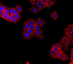
- Submitted by
- R&D Systems (provider)
- Main image
- Experimental details
- ER beta/NR3A2 in LNCaP Human Cell Line. ER beta/NR3A2 was detected in immersion fixed LNCaP human prostate cancer cell line using Mouse Anti-Human ER beta/NR3A2 Monoclonal Antibody (Catalog # MAB7106) at 10 µg/mL for 3 hours at room temperature. Cells were stained using the NorthernLights™ 557-conjugated Anti-Mouse IgG Secondary Antibody (red; Catalog # NL007) and counter-stained with DAPI (blue). Specific staining was localized to cytoplasm. View our protocol for Fluorescent ICC Staining of Cells on Coverslips.

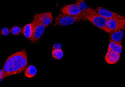
- Submitted by
- R&D Systems (provider)
- Main image
- Experimental details
- ER beta/NR3A2 in MCF-7 Human Cell Line. ER beta/NR3A2 was detected in immersion fixed MCF-7 human breast cancer cell line using Mouse Anti-Human ER beta/NR3A2 Monoclonal Antibody (Catalog # MAB7106) at 10 µg/mL for 3 hours at room temperature. Cells were stained using the NorthernLights™ 557-conjugated Anti-Mouse IgG Secondary Antibody (red; Catalog # NL007) and counter-stained with DAPI (blue). Specific staining was localized to cytoplasm and nuclei. View our protocol for Fluorescent ICC Staining of Cells on Coverslips.

Supportive validation
- Submitted by
- R&D Systems (provider)
- Main image
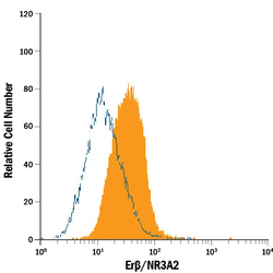
- Experimental details
- Detection of ER beta/NR3A2 in MCF-7 Human Cell Line by Flow Cytometry. MCF-7 human breast cancer cell line was stained with Mouse Anti-Human ER beta/NR3A2 Monoclonal Antibody (Catalog # MAB7106, filled histogram) or isotype control antibody (Catalog # MAB002, open histogram), followed by Phycoerythrin-conjugated Anti-Mouse IgG Secondary Antibody (Catalog # F0102B). To facilitate intracellular staining, cells were fixed with paraformaldehyde and permeabilized with saponin.
