Explore
 Validate
 Learn
Learn Western blot
 Immunohistochemistry
Antibody data
- Antibody Data
- Antigen structure
- References [0]
- Comments [0]
- Validations
- Western blot [1]
- Immunocytochemistry [1]
- Immunohistochemistry [9]
Submit
Validation data
Reference
Comment
Report error
- Product number
- HPA054962 - Provider product page

- Provider
- Atlas Antibodies
- Proper citation
- Atlas Antibodies Cat#HPA054962, RRID:AB_2682652
- Product name
- Anti-RBL1
- Antibody type
- Polyclonal
- Reactivity
- Human
- Host
- Rabbit
- Conjugate
- Unconjugated
- Antigen sequence
ESLAWSHDSALWEALQVSANKVPTCEEVIFPNNFE
TGNGGNVQGHLPLMPMSPLMHPRVKEVRTDSGSLR
RDMQPLSPISVHER- Isotype
- IgG
- Vial size
- 100 µl
- Storage
- Store at +4°C for short term storage. Long time storage is recommended at -20°C.
No comments: Submit comment
Supportive validation
- Submitted by
- Atlas Antibodies (provider)
- Main image

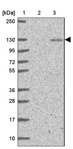
- Experimental details
- Lane 1: Marker [kDa] 250, 130, 95, 72, 55, 36, 28, 17, 10Lane 2: Human cell line RT-4Lane 3: Human cell line U-251MG sp
Supportive validation
- Submitted by
- Atlas Antibodies (provider)
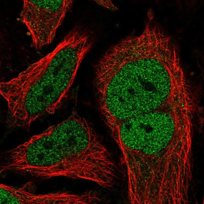
- Main image

- Experimental details
- Immunofluorescent staining of human cell line HeLa shows localization to nucleoplasm.
- Sample type
- HUMAN
Enhanced validation
Enhanced validation
Supportive validation
- Submitted by
- Atlas Antibodies (provider)
- Enhanced method
- Orthogonal validation
- Main image
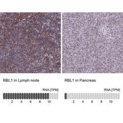
- Experimental details
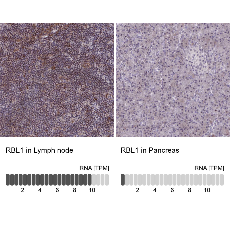
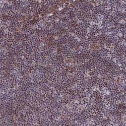
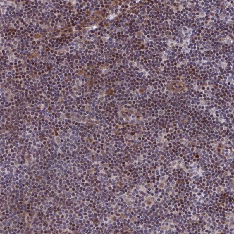
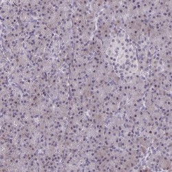
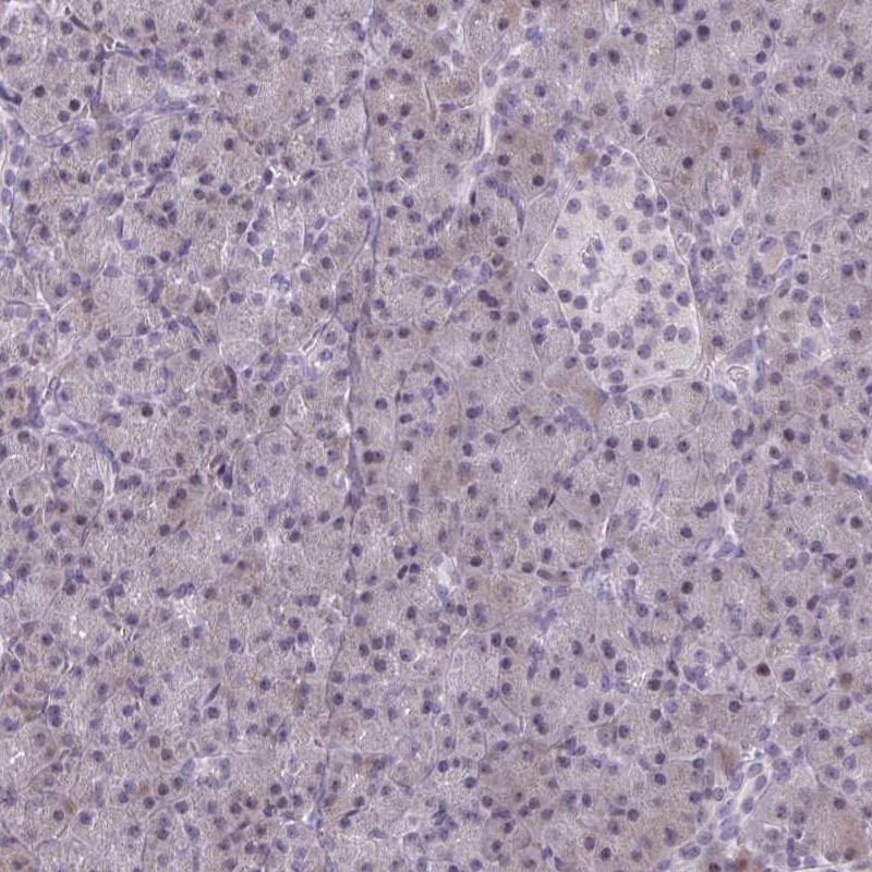
- Immunohistochemistry analysis in human lymph node and pancreas tissues using Anti-RBL1 antibody. Corresponding RBL1 RNA-seq data are presented for the same tissues.
- Sample type
- HUMAN
Enhanced validation
- Submitted by
- Atlas Antibodies (provider)
- Enhanced method
- Independent antibody validation
- Main image

- Experimental details
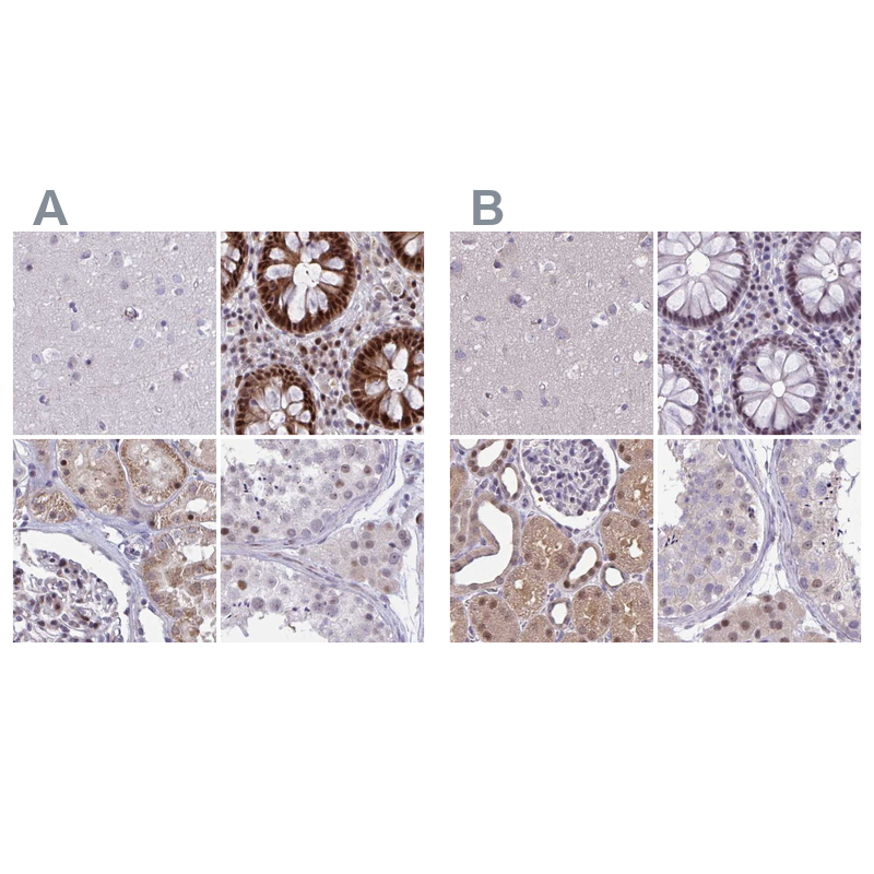
- Immunohistochemical staining of human cerebral cortex, colon, kidney and testis using Anti-RBL1 antibody HPA054962 (A) shows similar protein distribution across tissues to independent antibody HPA056525 (B).
Supportive validation
- Submitted by
- Atlas Antibodies (provider)
- Main image
- Experimental details
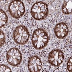
- Immunohistochemical staining of human colon shows strong nuclear and cytoplasmic positivity in glandular cells.
- Submitted by
- Atlas Antibodies (provider)
- Main image
- Experimental details
- Immunohistochemical staining of human lymph node shows high expression.
- Sample type
- HUMAN
- Submitted by
- Atlas Antibodies (provider)
- Main image
- Experimental details
- Immunohistochemical staining of human pancreas shows low expression as expected.
- Sample type
- HUMAN
- Submitted by
- Atlas Antibodies (provider)
- Main image

- Experimental details
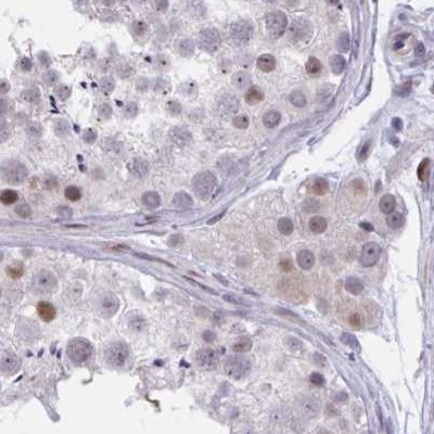
- Immunohistochemical staining of human testis using Anti-RBL1 antibody HPA054962.
- Sample type
- HUMAN
- Submitted by
- Atlas Antibodies (provider)
- Main image
- Experimental details
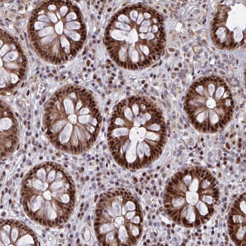
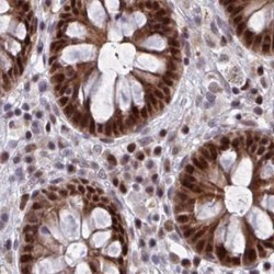
- Immunohistochemical staining of human colon using Anti-RBL1 antibody HPA054962.
- Sample type
- HUMAN

- Submitted by
- Atlas Antibodies (provider)
- Main image
- Experimental details
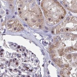
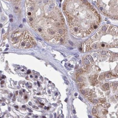
- Immunohistochemical staining of human kidney using Anti-RBL1 antibody HPA054962.
- Sample type
- HUMAN
- Submitted by
- Atlas Antibodies (provider)
- Main image
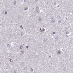
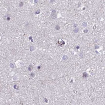
- Experimental details
- Immunohistochemical staining of human cerebral cortex using Anti-RBL1 antibody HPA054962.
- Sample type
- HUMAN