 Explore
 Validate
 Learn
Learn Western blot
 Immunocytochemistry
Antibody data
- Antibody Data
- Antigen structure
- References [1]
- Comments [0]
- Validations
- Western blot [1]
- Immunohistochemistry [1]
Submit
Validation data
Reference
Comment
Report error
- Product number
- NB100-2385 - Provider product page

- Provider
- Novus Biologicals
- Proper citation
- Novus Cat#NB100-2385, RRID:AB_10002905
- Product name
- Mouse Monoclonal S-arrestin Antibody
- Antibody type
- Monoclonal
- Description
- Protein G purified.
- Reactivity
- Human, Bovine, Porcine
- Host
- Mouse
- Isotype
- IgG
- Vial size
- 0.1 ml
- Concentration
- 1 mg/ml
- Storage
- Aliquot and store at -20C or -80C. Avoid freeze-thaw cycles.
Submitted references Immunoaffinity purification of S-antigen using monoclonal antibodies to different antigenic sites.
Banga JP, Suleyman S, Kasp E, Brown E, LeRoy F, Sanders M, Dumonde D
Investigative ophthalmology & visual science 1987 Mar;28(3):604-7
Investigative ophthalmology & visual science 1987 Mar;28(3):604-7
No comments: Submit comment
Supportive validation
- Submitted by
- Novus Biologicals (provider)
- Main image

- Experimental details
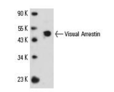
- Western Blot: S-arrestin Antibody (PDS-1) [NB100-2385] - Western blot analysis of Visual Arrestin expression in human retina tissue extract.
Supportive validation
- Submitted by
- Novus Biologicals (provider)
- Main image

- Experimental details
- Immunohistochemistry-Paraffin: S-arrestin Antibody (PDS-1) [NB100-2385] - S-arrestin was detected in immersion fixed paraffin-embedded sections of human prostate cancer using Mouse Anti-Human S-arrestin (PDS-1) Monoclonal Antibody (Catalog # NB100-2385) at 1:300 for 1 hour at room temperature followed by incubation with the Anti-Mouse IgG VisUCyte™ HRP Polymer Antibody (Catalog # VC001). Tissue was stained using DAB (brown) and counterstained with hematoxylin (blue). Specific staining was localized to the cytoplasm.
