 Explore
 Validate
 Learn
Learn Western blot
Antibody data
- Antibody Data
- Antigen structure
- References [1]
- Comments [0]
- Validations
- Western blot [1]
- Immunohistochemistry [9]
- Other assay [1]
Submit
Validation data
Reference
Comment
Report error
- Product number
- PA5-115530 - Provider product page

- Provider
- Invitrogen Antibodies
- Product name
- MACC1 Polyclonal Antibody
- Antibody type
- Polyclonal
- Antigen
- Synthetic peptide
- Reactivity
- Human, Mouse, Rat
- Host
- Rabbit
- Isotype
- IgG
- Vial size
- 100 µL
- Concentration
- 1 mg/mL
- Storage
- -20°C
Submitted references miR-199b-3p contributes to acquired resistance to cetuximab in colorectal cancer by targeting CRIM1 via Wnt/β-catenin signaling.
Han H, Li Y, Qin W, Wang L, Yin H, Su B, Yuan X
Cancer cell international 2022 Jan 28;22(1):42
Cancer cell international 2022 Jan 28;22(1):42
No comments: Submit comment
Supportive validation
- Submitted by
- Invitrogen Antibodies (provider)
- Main image

- Experimental details
- Western blot analysis of MACC1 in SW60 cells (left lane: treated with blocking peptide). Samples were incubated with polyclonal antibody (Product # PA5-115530).

Supportive validation
- Submitted by
- Invitrogen Antibodies (provider)
- Main image

- Experimental details
- Immunohistochemistry analysis of MACC1 in rat heart tissue. Samples were treated with formaldehyde and treated with citrate buffer for antigen retrieval, blocked, and incubated (1.5 hours at 22°C) with polyclonal antibody (Product # PA5-115530) at a dilution of 1:100. Secondary staining was applied with HRP conjugated anti-Rabbit.

- Submitted by
- Invitrogen Antibodies (provider)
- Main image

- Experimental details
- Immunohistochemistry analysis of MACC1 in mouse kidney tissue. Samples were treated with formaldehyde and treated with citrate buffer for antigen retrieval, blocked, and incubated (1.5 hours at 22°C) with polyclonal antibody (Product # PA5-115530) at a dilution of 1:100. Secondary staining was applied with HRP conjugated anti-Rabbit.

- Submitted by
- Invitrogen Antibodies (provider)
- Main image
- Experimental details
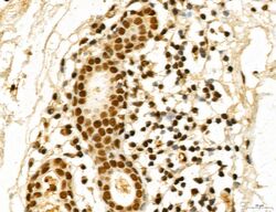
- Immunohistochemistry analysis of MACC1 in human normal tissues adjacent to esophageal cancer. Samples were treated with formaldehyde and treated with citrate buffer for antigen retrieval, blocked, and incubated (1.5 hours at 22°C) with polyclonal antibody (Product # PA5-115530) at a dilution of 1:100. Secondary staining was applied with HRP conjugated anti-Rabbit.

- Submitted by
- Invitrogen Antibodies (provider)
- Main image

- Experimental details
- Immunohistochemistry analysis of MACC1 in human esophageal cancer. Samples were treated with formaldehyde and treated with citrate buffer for antigen retrieval, blocked, and incubated (1.5 hours at 22°C) with polyclonal antibody (Product # PA5-115530) at a dilution of 1:100. Secondary staining was applied with HRP conjugated anti-Rabbit.

- Submitted by
- Invitrogen Antibodies (provider)
- Main image

- Experimental details
- Immunohistochemistry analysis of MACC1 in human colorectal cancer. Samples were treated with formaldehyde and treated with citrate buffer for antigen retrieval, blocked, and incubated (1.5 hours at 22°C) with polyclonal antibody (Product # PA5-115530) at a dilution of 1:100. Secondary staining was applied with HRP conjugated anti-Rabbit.

- Submitted by
- Invitrogen Antibodies (provider)
- Main image

- Experimental details
- Immunohistochemistry analysis of MACC1 in human mammary cancer. Samples were treated with formaldehyde and treated with citrate buffer for antigen retrieval, blocked, and incubated (1.5 hours at 22°C) with polyclonal antibody (Product # PA5-115530) at a dilution of 1:100. Secondary staining was applied with HRP conjugated anti-Rabbit.

- Submitted by
- Invitrogen Antibodies (provider)
- Main image

- Experimental details
- Immunohistochemistry analysis of MACC1 in human lung cancer. Samples were treated with formaldehyde and treated with citrate buffer for antigen retrieval, blocked, and incubated (1.5 hours at 22°C) with polyclonal antibody (Product # PA5-115530) at a dilution of 1:100. Secondary staining was applied with HRP conjugated anti-Rabbit.

- Submitted by
- Invitrogen Antibodies (provider)
- Main image

- Experimental details
- Immunohistochemistry analysis of MACC1 in rat kidney tissue. Samples were treated with formaldehyde and treated with citrate buffer for antigen retrieval, blocked, and incubated (1.5 hours at 22°C) with polyclonal antibody (Product # PA5-115530) at a dilution of 1:100. Secondary staining was applied with HRP conjugated anti-Rabbit.

- Submitted by
- Invitrogen Antibodies (provider)
- Main image

- Experimental details
- Immunohistochemistry analysis of MACC1 in rat ovary tissue. Samples were treated with formaldehyde and treated with citrate buffer for antigen retrieval, blocked, and incubated (1.5 hours at 22°C) with polyclonal antibody (Product # PA5-115530) at a dilution of 1:100. Secondary staining was applied with HRP conjugated anti-Rabbit.

Supportive validation
- Submitted by
- Invitrogen Antibodies (provider)
- Main image

- Experimental details
- Fig. 6 AntagomiR-199b-3p enhances the efficacy of CTx on CRC-CTxR tumor growth in vivo. After cell inoculation for a week, the volume of xenograft tumors in each group was recorded every five days (days 7, 12, 17, 22, 27, 32, and 37), A Growth curve and B representative images of xenograft tumors from each group. Tumor weights and D volume were recorded at the end of the experiments. E IHC staining determined the protein expression of Ki-67 and MACC1 in xenograft tumors (Scale bars: 100 mum). *P < 0.05 and ***P < 0.005 compared with saline group; ### P < 0.005 compared with CTx group
