 Explore
 Validate
 Learn
LearnHPA006504
antibody from Atlas Antibodies
Targeting: MIS18BP1
C14orf106, FLJ11186, KIAA1903, KNL2, M18BP1
 Western blot
Antibody data
- Antibody Data
- Antigen structure
- References [0]
- Comments [0]
- Validations
- Western blot [1]
- Immunocytochemistry [2]
- Immunohistochemistry [5]
Submit
Validation data
Reference
Comment
Report error
- Product number
- HPA006504 - Provider product page

- Provider
- Atlas Antibodies
- Proper citation
- Atlas Antibodies Cat#HPA006504, RRID:AB_1078317
- Product name
- Anti-MIS18BP1
- Antibody type
- Polyclonal
- Description
- Polyclonal Antibody against Human MIS18BP1, Gene description: MIS18 binding protein 1, Alternative Gene Names: C14orf106, FLJ11186, KIAA1903, KNL2, M18BP1, Validated applications: ICC, IHC, Uniprot ID: Q6P0N0, Storage: Store at +4°C for short term storage. Long time storage is recommended at -20°C.
- Reactivity
- Human
- Host
- Rabbit
- Conjugate
- Unconjugated
- Isotype
- IgG
- Vial size
- 100 µl
- Concentration
- 0.1 mg/ml
- Storage
- Store at +4°C for short term storage. Long time storage is recommended at -20°C.
No comments: Submit comment
Supportive validation
- Submitted by
- Atlas Antibodies (provider)
- Main image
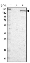
- Experimental details
- Lane 1: Marker [kDa] 230, 130, 95, 72, 56, 36, 28, 17, 11Lane 2: Human cell line RT-4Lane 3: Human cell line U-251MG sp

Supportive validation
- Submitted by
- Atlas Antibodies (provider)
- Main image

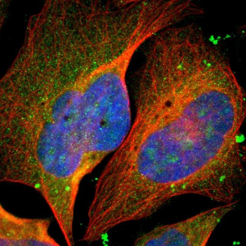
- Experimental details
- Immunofluorescent staining of human cell line U-2 OS shows localization to nucleoli & cytosol.
- Sample type
- HUMAN
- Submitted by
- Atlas Antibodies (provider)
- Main image

- Experimental details
- Immunofluorescent staining of human cell line U-2 OS shows localization to nucleoli & cytosol.
- Sample type
- Human
- Protocol
- Protocol

Supportive validation
- Submitted by
- Atlas Antibodies (provider)
- Main image
- Experimental details
- Immunohistochemical staining of human spleen shows moderate nuclear positivity in cells in red and white pulp.
- Sample type
- HUMAN

- Submitted by
- Atlas Antibodies (provider)
- Main image

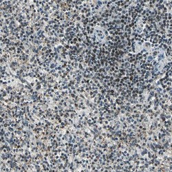
- Experimental details
- Immunohistochemical staining of human lymphoid node shows strong nuclear positivity in non-germinal center cells.
- Sample type
- Human
- Protocol
- Protocol

- Submitted by
- Atlas Antibodies (provider)
- Main image

- Experimental details
- Immunohistochemical staining of human testis shows strong nuclear positivity in Leydig cells.
- Sample type
- Human
- Protocol
- Protocol

- Submitted by
- Atlas Antibodies (provider)
- Main image

- Experimental details
- Immunohistochemical staining of human rectum shows strong nuclear positivity in glandular cells.
- Sample type
- Human
- Protocol
- Protocol

- Submitted by
- Atlas Antibodies (provider)
- Main image

- Experimental details
- Immunohistochemical staining of human cerebral cortex shows strong nuclear positivity in neurons.
- Sample type
- Human
- Protocol
- Protocol
