Antibody data
- Antibody Data
- Antigen structure
- References [3]
- Comments [0]
- Validations
- Western blot [1]
Submit
Validation data
Reference
Comment
Report error
- Product number
- PAB9953 - Provider product page

- Provider
- Abnova Corporation
- Proper citation
- Abnova Corporation Cat#PAB9953, RRID:AB_1676781
- Product name
- IL6 polyclonal antibody
- Antibody type
- Polyclonal
- Antigen
- Recombinant protein corresponding to human IL6.
- Reactivity
- Human
- Host
- Rabbit
- Vial size
- 1 mL
- Storage
- Store at 4°C. For long term storage store at -20°C.Aliquot to avoid repeated freezing and thawing.
Submitted references Expression of TGF-beta isoforms by Thy-1+ and Thy-1- pulmonary fibroblast subsets: evidence for TGF-beta as a regulator of IL-1-dependent stimulation of IL-6.
Enhancement of interleukin 6 cytostatic effect on human breast carcinoma cells by soluble IL-6 receptor from urine and reversion by monoclonal antibody.
Monoclonal antibodies for affinity purification of IL-6/IFN- beta 2 and for neutralization of HGF activity.
Silvera MR, Sempowski GD, Phipps RP
Lymphokine and cytokine research 1994 Oct;13(5):277-85
Lymphokine and cytokine research 1994 Oct;13(5):277-85
Enhancement of interleukin 6 cytostatic effect on human breast carcinoma cells by soluble IL-6 receptor from urine and reversion by monoclonal antibody.
Novick D, Shulman LM, Chen L, Revel M
Cytokine 1992 Jan;4(1):6-11
Cytokine 1992 Jan;4(1):6-11
Monoclonal antibodies for affinity purification of IL-6/IFN- beta 2 and for neutralization of HGF activity.
Novick D, Eshhar Z, Revel M, Mory Y
Hybridoma 1989 Oct;8(5):561-7
Hybridoma 1989 Oct;8(5):561-7
No comments: Submit comment
Supportive validation
- Submitted by
- Abnova Corporation (provider)
- Main image

- Experimental details
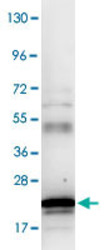
- Western blot using IL6 polyclonal antibody (Cat # PAB9953).Protein was resolved on a 4-20% Tris-Glycine gel by SDS-PAGE and transferred onto nitrocellulose. The blot shows detection of a band ~21 KDa in size corresponding to anti-IL6 antibody. After transfer, the membrane was blocked for 30 minutes with 1% BSA-TBST. Detection occurred using peroxidase conjugated anti-Rabbit IgG secondary antibody diluted 1 : 40,000 in blocking buffer for 30 min at RT followed by reaction with FemtoMax™ chemiluminescent substrate. Image was captured using VersaDoc™ MP 4000 imaging system (Bio-Rad).
- Validation comment
- Western Blot
 Explore
 Validate
 Learn
Learn Western blot
 ELISA
ELISA