OSM00017W
antibody from Invitrogen Antibodies
Targeting: MCOLN1
ML4, MLIV, MST080, MSTP080, TRPM-L1, TRPML1
Antibody data
- Antibody Data
- Antigen structure
- References [1]
- Comments [0]
- Validations
- Other assay [2]
Submit
Validation data
Reference
Comment
Report error
- Product number
- OSM00017W - Provider product page

- Provider
- Invitrogen Antibodies
- Product name
- TRPML1 Polyclonal Antibody
- Antibody type
- Polyclonal
- Antigen
- Synthetic peptide
- Description
- Excitation: 488-561 nm; Emission: 607 nm; Laser: Blue Laser, Green Laser, Yellow-Green Laser
- Reactivity
- Human, Mouse, Rat
- Host
- Rabbit
- Isotype
- IgG
- Vial size
- 100 µL
- Concentration
- Conc. Not Determined
- Storage
- Store at 4°C short term. For long term storage, store at -20°C, avoiding freeze/thaw cycles. Glycerol (1:1) may be added for added stability.
Submitted references MCOLN1 Promotes Proliferation and Predicts Poor Survival of Patients with Pancreatic Ductal Adenocarcinoma.
Hu ZD, Yan J, Cao KY, Yin ZQ, Xin WW, Zhang MF
Disease markers 2019;2019:9436047
Disease markers 2019;2019:9436047
No comments: Submit comment
Supportive validation
- Submitted by
- Invitrogen Antibodies (provider)
- Main image

- Experimental details
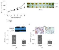
- Figure 3 In vivo animal experiments. (a) At the 14th day after injection, the tumor volume was measured every day. The growth curve showed that the tumors in the MCOLN1 group grew more slowly than controls. After inoculation of 29 days, the final tumor tissues were obtained. The tumors in the shRNA group were smaller than controls. (b, c) The expression of MCOLN1 in tumors of mice was detected by western blot or IHC. MCOLN1 expression was dramatically decreased in the shRNA group, which confirmed the effective silencing of MCOLN1 in tumors of mice in the shRNA group. ""*"" represents the significant association ( P < 0.05 for the difference was significant).
- Submitted by
- Invitrogen Antibodies (provider)
- Main image

- Experimental details
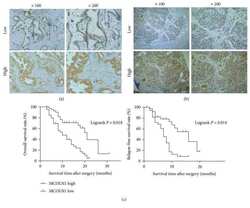
- Figure 4 The expression of MCOLN1 in tumor tissues and the associations of MCOLN1 with the prognosis of PDAC patients. (a, b) The staining of high and low expressions of MCOLN1 by IHC in PDAC tissues and paracarcinoma tissues. (c) OS and RFS rates with MCOLN1 expression in the 82 PDAC patients: shorter OS or RFS was significantly observed in high MCOLN1 expression than in low MCOLN1 expression in the 82 PDAC patients ( P < 0.05, respectively).
 Explore
 Validate
 Learn
Learn Western blot
 Other assay