Explore
 Validate
 Learn
LearnHPA031763
antibody from Atlas Antibodies
Targeting: MCOLN1
ML4, MLIV, MST080, MSTP080, TRPM-L1, TRPML1
 Western blot
 Immunohistochemistry
Antibody data
- Antibody Data
- Antigen structure
- References [0]
- Comments [0]
- Validations
- Western blot [3]
- Immunocytochemistry [1]
- Immunohistochemistry [6]
Submit
Validation data
Reference
Comment
Report error
- Product number
- HPA031763 - Provider product page

- Provider
- Atlas Antibodies
- Proper citation
- Atlas Antibodies Cat#HPA031763, RRID:AB_10600822
- Product name
- Anti-MCOLN1
- Antibody type
- Polyclonal
- Reactivity
- Human
- Host
- Rabbit
- Conjugate
- Unconjugated
- Antigen sequence
DTIKHPGGAGAEESELQAYIAQCQDSPTSGKFRRG
SGSACSLLCCCGRDPSE- Isotype
- IgG
- Vial size
- 100 µl
- Storage
- Store at +4°C for short term storage. Long time storage is recommended at -20°C.
No comments: Submit comment
Supportive validation
- Submitted by
- Atlas Antibodies (provider)
- Main image
- Experimental details
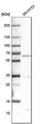
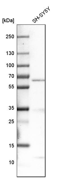
- Western blot analysis in human cell line SH-SY5Y.
- Sample type
- HUMAN
- Submitted by
- Atlas Antibodies (provider)
- Main image

- Experimental details
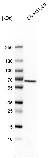
- Western blot analysis in human cell line SK-MEL-30.
- Sample type
- HUMAN
- Submitted by
- Atlas Antibodies (provider)
- Main image

- Experimental details
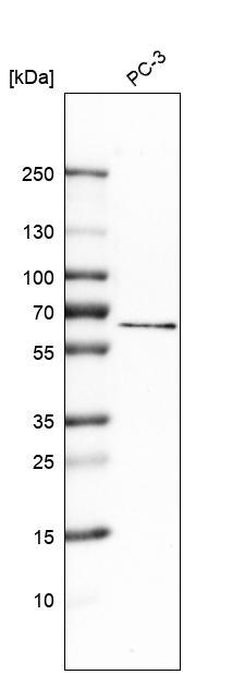
- Western blot analysis in human cell line PC-3.
Supportive validation
- Submitted by
- Atlas Antibodies (provider)
- Main image

- Experimental details
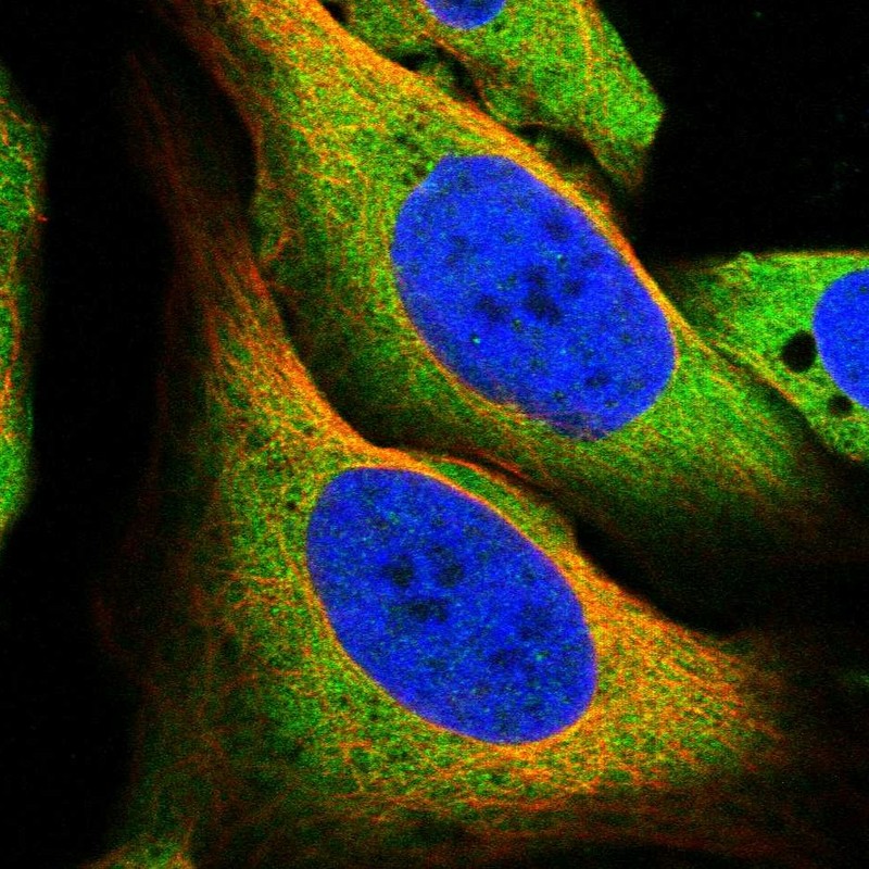
- Immunofluorescent staining of human cell line U-2 OS shows localization to plasma membrane & cytosol.
- Sample type
- HUMAN
Enhanced validation
Supportive validation
- Submitted by
- Atlas Antibodies (provider)
- Enhanced method
- Orthogonal validation
- Main image
- Experimental details
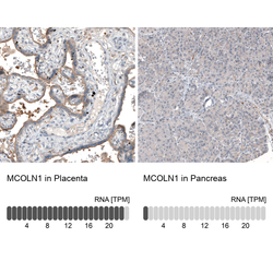
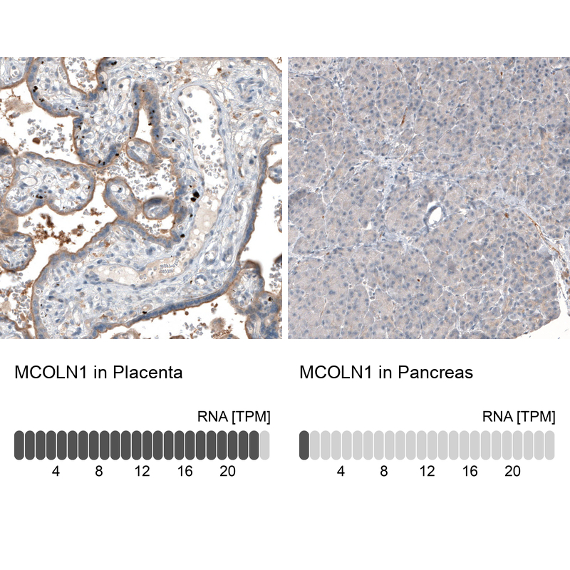
- Immunohistochemistry analysis in human placenta and pancreas tissues using HPA031763 antibody. Corresponding MCOLN1 RNA-seq data are presented for the same tissues.
- Sample type
- HUMAN
Supportive validation
- Submitted by
- Atlas Antibodies (provider)
- Main image
- Experimental details
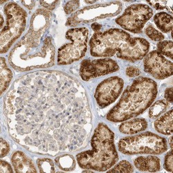
- Immunohistochemical staining of human kidney shows strong cytoplasmic positivity in cells in tubules.

- Submitted by
- Atlas Antibodies (provider)
- Main image

- Experimental details
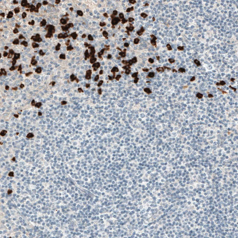
- Immunohistochemical staining of human tonsil shows strong cytoplasmic positivity in mast cell.
- Sample type
- HUMAN
- Submitted by
- Atlas Antibodies (provider)
- Main image

- Experimental details
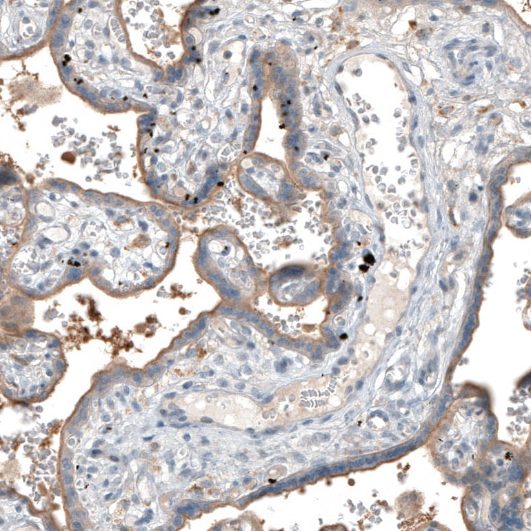
- Immunohistochemical staining of human placenta shows moderate cytoplasmic positivity in trophoblastic cells.
- Sample type
- HUMAN
- Submitted by
- Atlas Antibodies (provider)
- Main image
- Experimental details
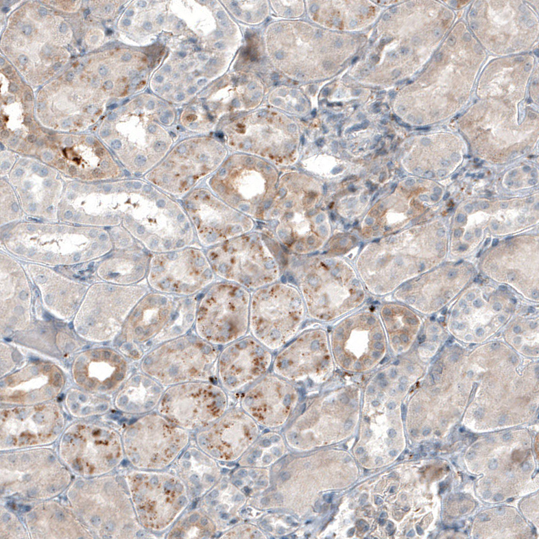
- Immunohistochemical staining of human kidney shows moderate granular cytoplasmic positivity in cells in tubules.
- Sample type
- HUMAN
- Submitted by
- Atlas Antibodies (provider)
- Main image
- Experimental details
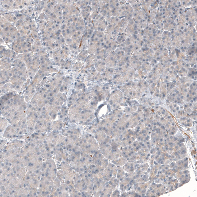
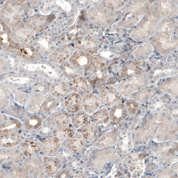
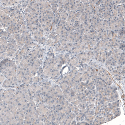
- Immunohistochemical staining of human pancreas shows no cytoplasmic positivity in endocrine glandular cells as expected.
- Sample type
- HUMAN