 Explore
 Validate
 Learn
Learn Western blot
Antibody data
- Antibody Data
- Antigen structure
- References [0]
- Comments [0]
- Validations
- Western blot [1]
- Immunocytochemistry [1]
- Immunohistochemistry [8]
Submit
Validation data
Reference
Comment
Report error
- Product number
- HPA023822 - Provider product page

- Provider
- Atlas Antibodies
- Proper citation
- Atlas Antibodies Cat#HPA023822, RRID:AB_1844982
- Product name
- Anti-ARFGEF1
- Antibody type
- Polyclonal
- Reactivity
- Human, Mouse
- Host
- Rabbit
- Conjugate
- Unconjugated
- Antigen sequence
QALQEAKQMEKERHRQHHHLLQSPVSHHEPESPQL
RYLPPQTVDHISQEHEGDLDLHTNDVDKSLQDDTE
PENGSDISSAENEQTEA- Isotype
- IgG
- Vial size
- 100 µl
- Storage
- Store at +4°C for short term storage. Long time storage is recommended at -20°C.
No comments: Submit comment
Supportive validation
- Submitted by
- Atlas Antibodies (provider)
- Main image

- Experimental details
- Lane 1: Marker [kDa] 250, 130, 100, 70, 55, 35, 25, 15, 10Lane 2: Human cell line RT-4

Supportive validation
- Submitted by
- Atlas Antibodies (provider)
- Main image

- Experimental details
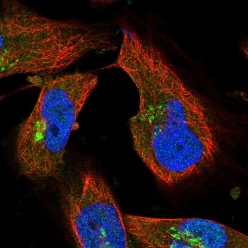
- Immunofluorescent staining of human cell line U-251 MG shows localization to nucleoplasm & the Golgi apparatus.
- Sample type
- HUMAN
Supportive validation
- Submitted by
- Atlas Antibodies (provider)
- Main image

- Experimental details
- Immunohistochemical staining of human cerebellum shows strong cytoplasmic positivity in Purkinje cells.
- Sample type
- HUMAN

- Submitted by
- Atlas Antibodies (provider)
- Main image

- Experimental details
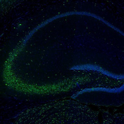
- Immunofluorescence staining of mouse hippocampus shows positivity in the CA3 area neurons.
- Sample type
- MOUSE
- Submitted by
- Atlas Antibodies (provider)
- Main image

- Experimental details
- Immunohistochemical staining of human cerebral cortex shows strong positivity in subsets of neurons.
- Sample type
- HUMAN

- Submitted by
- Atlas Antibodies (provider)
- Main image

- Experimental details
- Immunofluorescence staining of mouse olfactory bulb shows positivity in neurons and processes in the mitral and external plexiform layers.
- Sample type
- MOUSE

- Submitted by
- Atlas Antibodies (provider)
- Main image

- Experimental details
- Immunofluorescence staining of mouse piriform cortex shows neuronal staining in various layers.
- Sample type
- MOUSE

- Submitted by
- Atlas Antibodies (provider)
- Main image

- Experimental details
- Immunofluorescence staining of mouse parietal association cortex shows positivity in neuronal cell bodies and dendrites.
- Sample type
- MOUSE

- Submitted by
- Atlas Antibodies (provider)
- Main image

- Experimental details
- Immunofluorescence staining of mouse gigantocellular reticular nucleus shows immunoreactivity in neuronal cell bodies and processes.
- Sample type
- MOUSE

- Submitted by
- Atlas Antibodies (provider)
- Main image

- Experimental details
- Immunohistochemical staining of human cerebral cortex shows cytoplasmic immunoreactivity in neuronal cell bodies.
- Sample type
- HUMAN
