 Explore
 Validate
 Learn
Learn Western blot
 Immunocytochemistry
 Immunoprecipitation
Antibody data
- Antibody Data
- Antigen structure
- References [0]
- Comments [0]
- Validations
- Immunocytochemistry [1]
- Flow cytometry [4]
Submit
Validation data
Reference
Comment
Report error
- Product number
- MA1-16577 - Provider product page

- Provider
- Invitrogen Antibodies
- Product name
- BUBR1 Monoclonal Antibody (8G1)
- Antibody type
- Monoclonal
- Antigen
- Other
- Description
- Suggested positive control: Hela whole cell extract, antigen standard for BUB1B (transient overexpression lysate).
- Reactivity
- Human, Mouse, Rat
- Host
- Mouse
- Isotype
- IgG
- Antibody clone number
- 8G1
- Vial size
- 100 μL
- Concentration
- 1 mg/mL
- Storage
- -20°C, Avoid Freeze/Thaw Cycles
No comments: Submit comment
Supportive validation
- Submitted by
- Invitrogen Antibodies (provider)
- Main image

- Experimental details
- Immunofluorescence of an asynchronous cycling population of human cells (U2OS) with MA1-16577. No signal is detected from interphase cells, whereas cells undergoing mitosis accumulate BubR1 at the kinetochores. Image reveals kinetochores at prometaphase.

Supportive validation
- Submitted by
- Invitrogen Antibodies (provider)
- Main image

- Experimental details
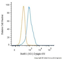
- Flow cytometry of BUBR1 in HeLa cells. Samples were incubated in BUBR1 monoclonal antibody (Product # MA1-16577) using a dilution of 5 µg/mL for 30 minutes at room temperature. Antibody (blue) and a matched isotype control (orange). Cells were fixed with 4% PFA and then permeabilized with 0.1% saponin. Both antibodies were conjugated to Dylight 650.

- Submitted by
- Invitrogen Antibodies (provider)
- Main image

- Experimental details
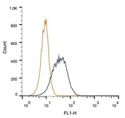
- Flow cytometry of BUBR1 in 1 x 10^6 HEK-293 cells. Samples were incubated in BUBR1 monoclonal antibody (Product # MA1-16577) using a dilution of 1 µg/1x10^6 cells. Antibody (dark blue). Isotype control shown in orange.

- Submitted by
- Invitrogen Antibodies (provider)
- Main image
- Experimental details
- Flow cytometry of BUBR1 in HeLa cells. Samples were incubated in BUBR1 monoclonal antibody (Product # MA1-16577) using a dilution of 5 µg/mL for 30 minutes at room temperature. Antibody (blue) and a matched isotype control (orange). Cells were fixed with 4% PFA and then permeabilized with 0.1% saponin. Both antibodies were conjugated to Dylight 650.

- Submitted by
- Invitrogen Antibodies (provider)
- Main image
- Experimental details
- Flow cytometry of BUBR1 in 1 x 10^6 HEK-293 cells. Samples were incubated in BUBR1 monoclonal antibody (Product # MA1-16577) using a dilution of 1 µg/1x10^6 cells. Antibody (dark blue). Isotype control shown in orange.
