Explore
 Validate
 Learn
Learn Western blot
 Immunocytochemistry
Antibody data
- Antibody Data
- Antigen structure
- References [9]
- Comments [0]
- Validations
- Western blot [1]
- Flow cytometry [2]
Submit
Validation data
Reference
Comment
Report error
- Product number
- NB100-353 - Provider product page

- Provider
- Novus Biologicals
- Proper citation
- Novus Cat#NB100-353, RRID:AB_10001219
- Product name
- Mouse Monoclonal BubR1 Antibody
- Antibody type
- Monoclonal
- Description
- Protein G purified. NB100-353 is specific for hBUBR1. There should be no cross-reactivity with hBUB1.
- Reactivity
- Human, Mouse
- Host
- Mouse
- Isotype
- IgG
- Vial size
- 0.1 ml
- Concentration
- 1.0 mg/ml
- Storage
- Aliquot and store at -20C or -80C. Avoid freeze-thaw cycles.
Submitted references Elucidating the Role of the Maternal Embryonic Leucine Zipper Kinase in Adrenocortical Carcinoma.
PKCε Controls Mitotic Progression by Regulating Centrosome Migration and Mitotic Spindle Assembly.
Plk1 is upregulated in androgen-insensitive prostate cancer cells and its inhibition leads to necroptosis.
Phosphoinositide 3-kinase β regulates chromosome segregation in mitosis.
A high-content, cell-based screen identifies micropolyin, a new inhibitor of microtubule dynamics.
Gradual reduction of BUBR1 protein levels results in premature sister-chromatid separation then in aneuploidy.
Dido disruption leads to centrosome amplification and mitotic checkpoint defects compromising chromosome stability.
Human BUBR1 is a mitotic checkpoint kinase that monitors CENP-E functions at kinetochores and binds the cyclosome/APC.
Human BUBR1 is a mitotic checkpoint kinase that monitors CENP-E functions at kinetochores and binds the cyclosome/APC.
Kiseljak-Vassiliades K, Zhang Y, Kar A, Razzaghi R, Xu M, Gowan K, Raeburn CD, Albuja-Cruz M, Jones KL, Somerset H, Fishbein L, Leong S, Wierman ME
Endocrinology 2018 Jul 1;159(7):2532-2544
Endocrinology 2018 Jul 1;159(7):2532-2544
PKCε Controls Mitotic Progression by Regulating Centrosome Migration and Mitotic Spindle Assembly.
Martini S, Soliman T, Gobbi G, Mirandola P, Carubbi C, Masselli E, Pozzi G, Parker PJ, Vitale M
Molecular cancer research : MCR 2018 Jan;16(1):3-15
Molecular cancer research : MCR 2018 Jan;16(1):3-15
Plk1 is upregulated in androgen-insensitive prostate cancer cells and its inhibition leads to necroptosis.
Deeraksa A, Pan J, Sha Y, Liu XD, Eissa NT, Lin SH, Yu-Lee LY
Oncogene 2013 Jun 13;32(24):2973-83
Oncogene 2013 Jun 13;32(24):2973-83
Phosphoinositide 3-kinase β regulates chromosome segregation in mitosis.
Silió V, Redondo-Muñoz J, Carrera AC
Molecular biology of the cell 2012 Dec;23(23):4526-42
Molecular biology of the cell 2012 Dec;23(23):4526-42
A high-content, cell-based screen identifies micropolyin, a new inhibitor of microtubule dynamics.
De Rycker M, Rigoreau L, Dowding S, Parker PJ
Chemical biology & drug design 2009 Jun;73(6):599-610
Chemical biology & drug design 2009 Jun;73(6):599-610
Gradual reduction of BUBR1 protein levels results in premature sister-chromatid separation then in aneuploidy.
Bohers E, Sarafan-Vasseur N, Drouet A, Paresy M, Latouche JB, Flaman JM, Sesboüé R, Frebourg T
Human genetics 2008 Dec;124(5):473-8
Human genetics 2008 Dec;124(5):473-8
Dido disruption leads to centrosome amplification and mitotic checkpoint defects compromising chromosome stability.
Trachana V, van Wely KH, Guerrero AA, Fütterer A, Martínez-A C
Proceedings of the National Academy of Sciences of the United States of America 2007 Feb 20;104(8):2691-6
Proceedings of the National Academy of Sciences of the United States of America 2007 Feb 20;104(8):2691-6
Human BUBR1 is a mitotic checkpoint kinase that monitors CENP-E functions at kinetochores and binds the cyclosome/APC.
Chan GK, Jablonski SA, Sudakin V, Hittle JC, Yen TJ
The Journal of cell biology 1999 Sep 6;146(5):941-54
The Journal of cell biology 1999 Sep 6;146(5):941-54
Human BUBR1 is a mitotic checkpoint kinase that monitors CENP-E functions at kinetochores and binds the cyclosome/APC.
Chan GK, Jablonski SA, Sudakin V, Hittle JC, Yen TJ
The Journal of cell biology 1999 Sep 6;146(5):941-54
The Journal of cell biology 1999 Sep 6;146(5):941-54
No comments: Submit comment
Supportive validation
- Submitted by
- Novus Biologicals (provider)
- Main image
- Experimental details
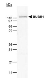
- Western Blot: BubR1 Antibody (8G1) [NB100-353] - BUBR1 detected in HeLa cell lysate using NB 100-353.

Supportive validation
- Submitted by
- Novus Biologicals (provider)
- Main image

- Experimental details
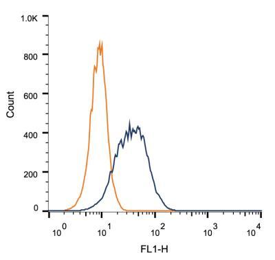
- Flow Cytometry: BubR1 Antibody (8G1) [NB100-353] - Intracellular flow cytometric staining of 1 x 10^6 HEK-293 cells using BubR1 antibody (dark blue). Isotype control shown in orange. An antibody concentration of 1 ug/1x10^6 cells was used.
- Submitted by
- Novus Biologicals (provider)
- Main image

- Experimental details
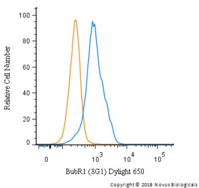
- Flow Cytometry: BubR1 Antibody (8G1) [NB100-353] - An intracellular stain was performed on HeLa cells with BubR1 Antibody (8G1)NB100-353C (blue) and a matched isotype control (orange). Cells were fixed with 4% PFA and then permeabilized with 0.1% saponin. Cells were incubated in an antibody dilution of 5 ug/mL for 30 minutes at room temperature. Both antibodies were conjugated to Dylight 650.