Explore
 Validate
 Learn
Learn Western blot
Antibody data
- Antibody Data
- Antigen structure
- References [0]
- Comments [0]
- Validations
- Western blot [2]
- Immunohistochemistry [1]
Submit
Validation data
Reference
Comment
Report error
- Product number
- AGR-013-200UL - Provider product page

- Provider
- Invitrogen Antibodies
- Product name
- GALR3 (extracellular) Polyclonal Antibody
- Antibody type
- Polyclonal
- Antigen
- Other
- Description
- Reconstitution: 1 X 50 µL double distilled water (DDW), depending on the sample size. The antibody ships as a lyophilized powder at room temperature. Upon arrival, it should be stored at -20C. The reconstituted solution can be stored at 4C for up to 1 week. For longer periods, small aliquots should be stored at -20C. Avoid multiple freezing and thawing. Centrifuge all antibody preparations before use (10000 x g 5 min).
- Reactivity
- Human, Mouse, Rat
- Host
- Rabbit
- Isotype
- IgG
- Vial size
- 200 µL
- Concentration
- 0.7 mg/mL
- Storage
- -20° C, Avoid Freeze/Thaw Cycles
No comments: Submit comment
Supportive validation
- Submitted by
- Invitrogen Antibodies (provider)
- Main image
- Experimental details
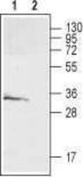
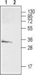
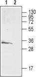
- Western blot analysis of rat brain membrane: - 1. Anti-GALR3 (extracellular) Antibody (#AGR-013), (1:200). 2. Anti-GALR3 (extracellular) Antibody , preincubated with GALR3 (extracellular) Blocking Peptide (#BLP-GR013).
- Submitted by
- Invitrogen Antibodies (provider)
- Main image
- Experimental details
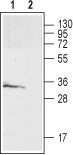
- Western blot analysis of rat brain membrane: - 1. Anti-GALR3 (extracellular) Antibody (#AGR-013), (1:200). 2. Anti-GALR3 (extracellular) Antibody , preincubated with GALR3 (extracellular) Blocking Peptide (#BLP-GR013).
Supportive validation
- Submitted by
- Invitrogen Antibodies (provider)
- Main image

- Experimental details
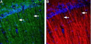
- Expression of Galanin receptor 3 in rat hippocampus - Immunohistochemical staining of rat hippocampus using Anti-GALR3 (extracellular) Antibody (#AGR-013). A. Galanin receptor 3 (green) appears in apical dendrites of pyramidal neurons in CA1 (arrows). B. MAP2 (red), a marker of axons and dendrites, is stained in the same section. GALR3 appears to be expressed in a subset of apical dendrites (arrows). DAPI is used as the counterstain (blue).