Explore
 Validate
 Learn
LearnPA5-115149
antibody from Invitrogen Antibodies
Targeting: DYNC1H1
CMT2O, DHC1, DNCH1, Dnchc1, DNCL, DNECL, HL-3, p22
 Western blot
Antibody data
- Antibody Data
- Antigen structure
- References [0]
- Comments [0]
- Validations
- Western blot [2]
- Immunocytochemistry [1]
- Immunohistochemistry [4]
Submit
Validation data
Reference
Comment
Report error
- Product number
- PA5-115149 - Provider product page

- Provider
- Invitrogen Antibodies
- Product name
- DYNC1H1 Polyclonal Antibody
- Antibody type
- Polyclonal
- Antigen
- Synthetic peptide
- Reactivity
- Human, Mouse, Rat
- Host
- Rabbit
- Isotype
- IgG
- Vial size
- 100 µL
- Concentration
- 1 mg/mL
- Storage
- -20°C
No comments: Submit comment
Supportive validation
- Submitted by
- Invitrogen Antibodies (provider)
- Main image
- Experimental details
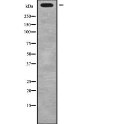
- Western blot analysis of DYNC1H1 in NIH-3T3 whole cells lysates. Samples were incubated with polyclonal antibody (Product # PA5-115149).

- Submitted by
- Invitrogen Antibodies (provider)
- Main image

- Experimental details
- Western blot analysis of DYNC1H1 in HepG2 and rat muscle and mouse brain. Samples were incubated with polyclonal antibody (Product # PA5-115149).

Supportive validation
- Submitted by
- Invitrogen Antibodies (provider)
- Main image
- Experimental details
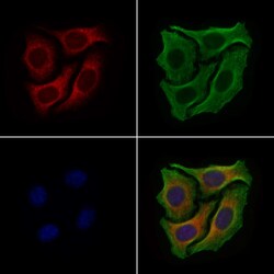
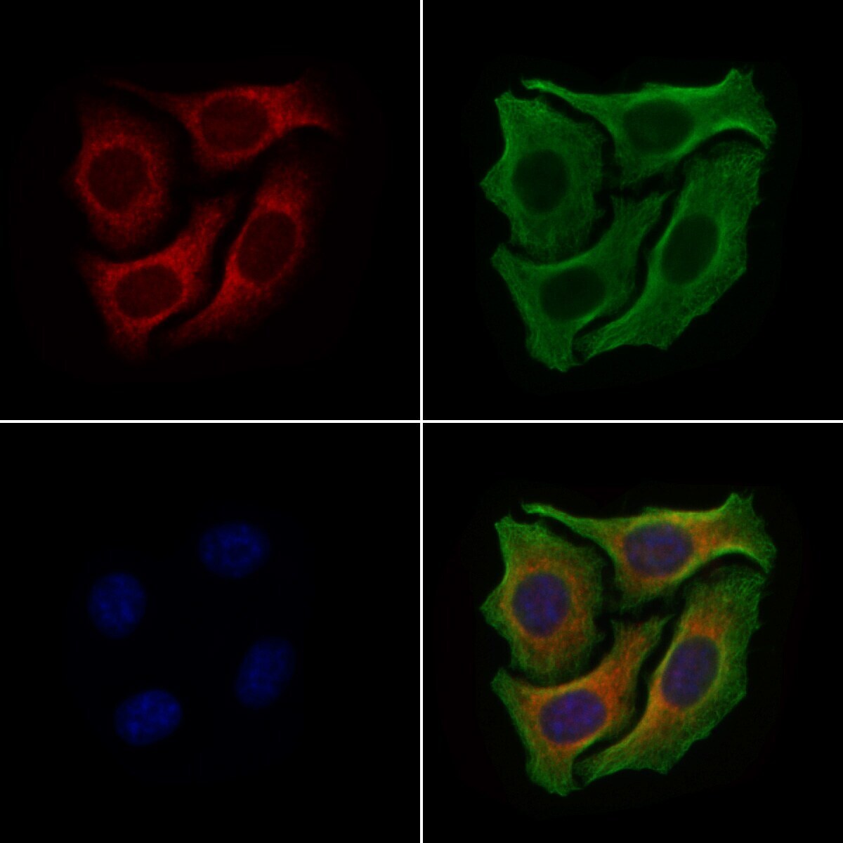
- Immunocytochemistry analysis of DYNC1H1 in HeLa cells. Samples were treated with PFA, permeabilized in 0.1% Triton X-100, blocked in 10% serum (45 min at 25°C), and incubated with polyclonal antibody (Product # PA5-115149) at a dilution of 1:200 (1 hr at 37°C). Secondary staining was applied with mouse anti-beta tubulin; AlexaFluor 594 conjugated goat anti-rabbit IgG (Red); AlexaFluor 88 conjugated goat anti-mouse IgG (Green) and DAPI (blue) using a dilution of 1:200 (1 hr at 37°C).
Supportive validation
- Submitted by
- Invitrogen Antibodies (provider)
- Main image

- Experimental details
- Immunohistochemistry analysis of DYNC1H1 in mouse kidney tissue. Samples were treated with formaldehyde and treated with citrate buffer for antigen retrieval, blocked, and incubated (1.5 hours at 22°C) with polyclonal antibody (Product # PA5-115149) at a dilution of 1:100. Secondary staining was applied with HRP conjugated goat anti-rabbit.

- Submitted by
- Invitrogen Antibodies (provider)
- Main image

- Experimental details
- Immunohistochemistry analysis of DYNC1H1 in human kidney cancer and adjacent normal tissues. Samples were treated with formaldehyde and treated with citrate buffer for antigen retrieval, blocked, and incubated (1.5 hours at 22°C) with polyclonal antibody (Product # PA5-115149) at a dilution of 1:100. Secondary staining was applied with HRP conjugated anti-Rabbit.

- Submitted by
- Invitrogen Antibodies (provider)
- Main image

- Experimental details
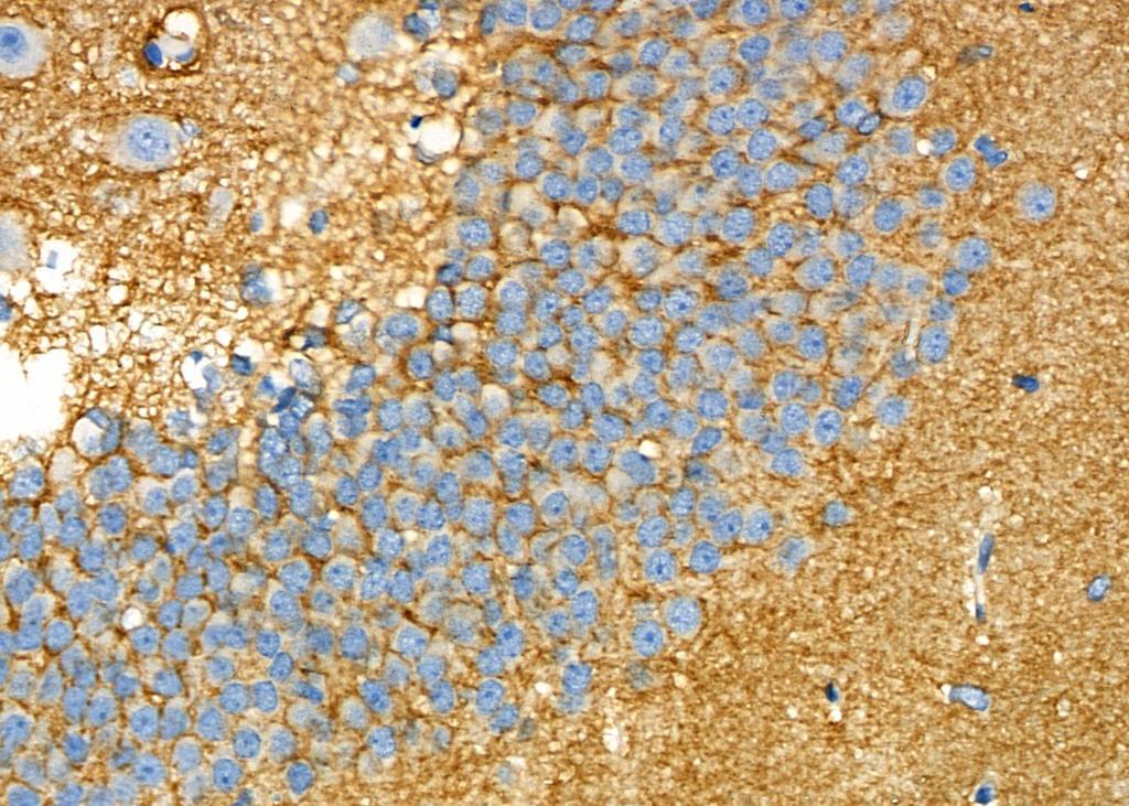
- Immunohistochemistry analysis of DYNC1H1 in mouse brain tissue. Samples were treated with formaldehyde and treated with citrate buffer for antigen retrieval, blocked, and incubated (1.5 hours at 22°C) with polyclonal antibody (Product # PA5-115149) at a dilution of 1:100. Secondary staining was applied with HRP conjugated anti-Rabbit.

- Submitted by
- Invitrogen Antibodies (provider)
- Main image

- Experimental details
- Immunohistochemistry analysis of DYNC1H1 in rat brain tissue. Samples were treated with formaldehyde and treated with citrate buffer for antigen retrieval, blocked, and incubated (1.5 hours at 22°C) with polyclonal antibody (Product # PA5-115149) at a dilution of 1:100. Secondary staining was applied with HRP conjugated anti-Rabbit.