Explore
 Validate
 Learn
Learn Western blot
Antibody data
- Antibody Data
- Antigen structure
- References [10]
- Comments [0]
- Validations
- Western blot [2]
- Immunoprecipitation [1]
- Immunohistochemistry [2]
- Flow cytometry [1]
- Other assay [12]
Submit
Validation data
Reference
Comment
Report error
- Product number
- 43-7800 - Provider product page

- Provider
- Invitrogen Antibodies
- Product name
- Caspase 3 Monoclonal Antibody (74T2)
- Antibody type
- Monoclonal
- Antigen
- Recombinant full-length protein
- Reactivity
- Human, Mouse, Rat
- Host
- Mouse
- Isotype
- IgG
- Antibody clone number
- 74T2
- Vial size
- 100 µg
- Concentration
- 0.5 mg/mL
- Storage
- Store at 4°C short term. For long term storage, store at -20°C, avoiding freeze/thaw cycles.
Submitted references 2-Methoxyestradiol combined with ascorbic acid facilitates the apoptosis of chronic myeloid leukemia cells via the microRNA-223/Fms-like tyrosine kinase 3/phosphatidylinositol-3 kinase/protein kinase B axis.
The Ganglioside Monosialotetrahexosylganglioside Protects Auditory Hair Cells Against Neomycin-Induced Cytotoxicity Through Mitochondrial Antioxidation: An in vitro Study.
Compatibility of ingredients of Danshen (Radix Salviae Miltiorrhizae) and Honghua (Flos Carthami) and their protective effects on cerebral ischemia-reperfusion injury in rats.
Antibody-drug nanoparticle induces synergistic treatment efficacies in HER2 positive breast cancer cells.
Iron oxides nanoparticles (IOs) exposed to magnetic field promote expression of osteogenic markers in osteoblasts through integrin alpha-3 (INTa-3) activation, inhibits osteoclasts activity and exerts anti-inflammatory action.
5-Azacytydine and resveratrol reverse senescence and ageing of adipose stem cells via modulation of mitochondrial dynamics and autophagy.
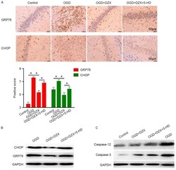
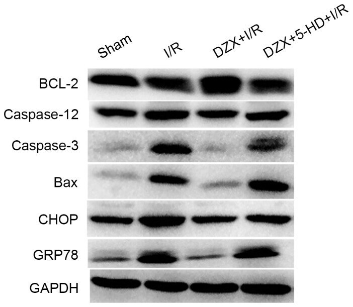
Diazoxide inhibits of ER stress‑mediated apoptosis during oxygen‑glucose deprivation in vitro and cerebral ischemia‑reperfusion in vivo.
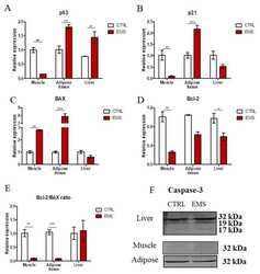
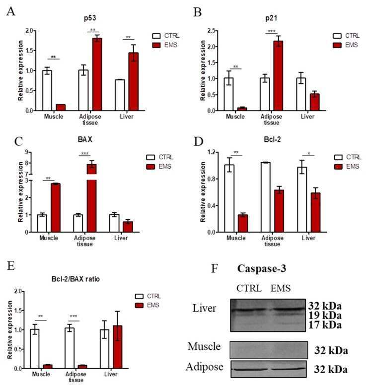
Excessive Endoplasmic Reticulum Stress Correlates with Impaired Mitochondrial Dynamics, Mitophagy and Apoptosis, in Liver and Adipose Tissue, but Not in Muscles in EMS Horses.
Ultrastructural changes during osteogenic differentiation in mesenchymal stromal cells cultured in alginate hydrogel.
Tunneling nanotubes: an alternate route for propagation of the bystander effect following oncolytic viral infection.
Zhang S, Yu H, Li J, Fan J, Chen J
Bioengineered 2022 Feb;13(2):3470-3485
Bioengineered 2022 Feb;13(2):3470-3485
The Ganglioside Monosialotetrahexosylganglioside Protects Auditory Hair Cells Against Neomycin-Induced Cytotoxicity Through Mitochondrial Antioxidation: An in vitro Study.
Li Y, Li A, Wang C, Jin X, Zhang Y, Lu L, Wang SL, Gao X
Frontiers in cellular neuroscience 2021;15:751867
Frontiers in cellular neuroscience 2021;15:751867
Compatibility of ingredients of Danshen (Radix Salviae Miltiorrhizae) and Honghua (Flos Carthami) and their protective effects on cerebral ischemia-reperfusion injury in rats.
Wan H, Yang Y, Li Z, Cheng L, Ding Z, Wan H, Yang J, Zhou H
Experimental and therapeutic medicine 2021 Aug;22(2):849
Experimental and therapeutic medicine 2021 Aug;22(2):849
Antibody-drug nanoparticle induces synergistic treatment efficacies in HER2 positive breast cancer cells.
Abedin MR, Powers K, Aiardo R, Barua D, Barua S
Scientific reports 2021 Apr 1;11(1):7347
Scientific reports 2021 Apr 1;11(1):7347
Iron oxides nanoparticles (IOs) exposed to magnetic field promote expression of osteogenic markers in osteoblasts through integrin alpha-3 (INTa-3) activation, inhibits osteoclasts activity and exerts anti-inflammatory action.
Marycz K, Sobierajska P, Roecken M, Kornicka-Garbowska K, Kępska M, Idczak R, Nedelec JM, Wiglusz RJ
Journal of nanobiotechnology 2020 Feb 18;18(1):33
Journal of nanobiotechnology 2020 Feb 18;18(1):33
5-Azacytydine and resveratrol reverse senescence and ageing of adipose stem cells via modulation of mitochondrial dynamics and autophagy.
Kornicka K, Szłapka-Kosarzewska J, Śmieszek A, Marycz K
Journal of cellular and molecular medicine 2019 Jan;23(1):237-259
Journal of cellular and molecular medicine 2019 Jan;23(1):237-259
Diazoxide inhibits of ER stress‑mediated apoptosis during oxygen‑glucose deprivation in vitro and cerebral ischemia‑reperfusion in vivo.
Lei X, Lei L, Zhang Z, Cheng Y
Molecular medicine reports 2018 Jun;17(6):8039-8046
Molecular medicine reports 2018 Jun;17(6):8039-8046
Excessive Endoplasmic Reticulum Stress Correlates with Impaired Mitochondrial Dynamics, Mitophagy and Apoptosis, in Liver and Adipose Tissue, but Not in Muscles in EMS Horses.
Marycz K, Kornicka K, Szlapka-Kosarzewska J, Weiss C
International journal of molecular sciences 2018 Jan 6;19(1)
International journal of molecular sciences 2018 Jan 6;19(1)
Ultrastructural changes during osteogenic differentiation in mesenchymal stromal cells cultured in alginate hydrogel.
Grzesiak J, Śmieszek A, Marycz K
Cell & bioscience 2017;7:2
Cell & bioscience 2017;7:2
Tunneling nanotubes: an alternate route for propagation of the bystander effect following oncolytic viral infection.
Ady J, Thayanithy V, Mojica K, Wong P, Carson J, Rao P, Fong Y, Lou E
Molecular therapy oncolytics 2016;3:16029
Molecular therapy oncolytics 2016;3:16029
No comments: Submit comment
Supportive validation
- Submitted by
- Invitrogen Antibodies (provider)
- Main image
- Experimental details
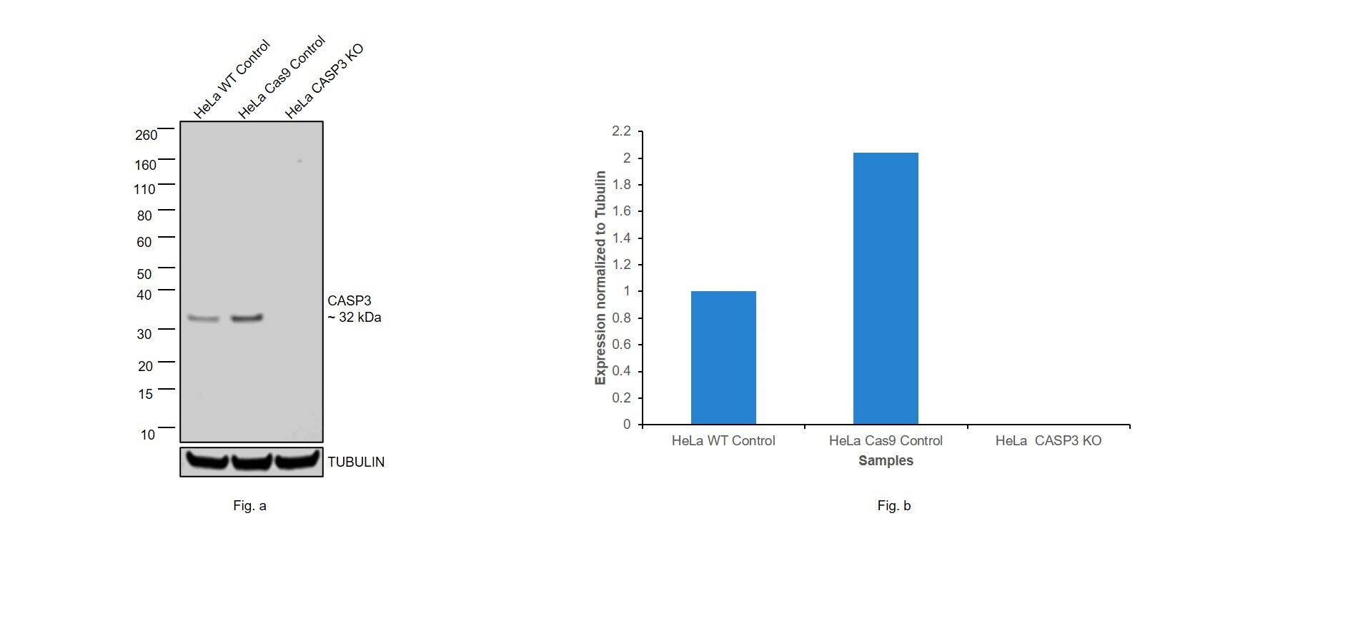
- Knockout of CASP3 was achieved by CRISPR-Cas9 genome editing using LentiArray™ Lentiviral sgRNA (Product # A32042, Assay ID CRISPR797315_LV) and LentiArray Cas9 Lentivirus (Product # A32064). Western blot analysis of CASP3 was performed by loading 50 µg of HeLa Wild Type (Lane 1), HeLa Cas9 (Lane 2) andHeLa CASP3 KO (Lane 3) whole cell extracts. The samples were electrophoresed using NuPAGE™ Novex™ 4-12% Bis-Tris Protein Gel (Product # NP0322BOX). Resolved proteins were then transferred onto a nitrocellulose membrane (Product # IB23001) by iBlot® 2 Dry Blotting System (Product # IB21001). The blot was probed with Anti-Caspase 3 Monoclonal Antibody (74T2) (Product # 43-7800, 1 µg/mL dilution) and Goat anti-Mouse IgG (H+L) Superclonal™ Recombinant Secondary Antibody, HRP (Product # A28177, 1:8,000 dilution) using the iBright FL 1000 (Product # A32752). Chemiluminescent detection was performed using Novex® ECL Chemiluminescent Substrate Reagent Kit (Product # WP20005). Loss of signal upon CRISPR mediated knockout (KO) using the LentiArray™ CRISPR product line confirms that antibody is specific to CASP3.
- Submitted by
- Invitrogen Antibodies (provider)
- Main image
- Experimental details
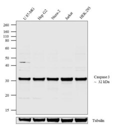
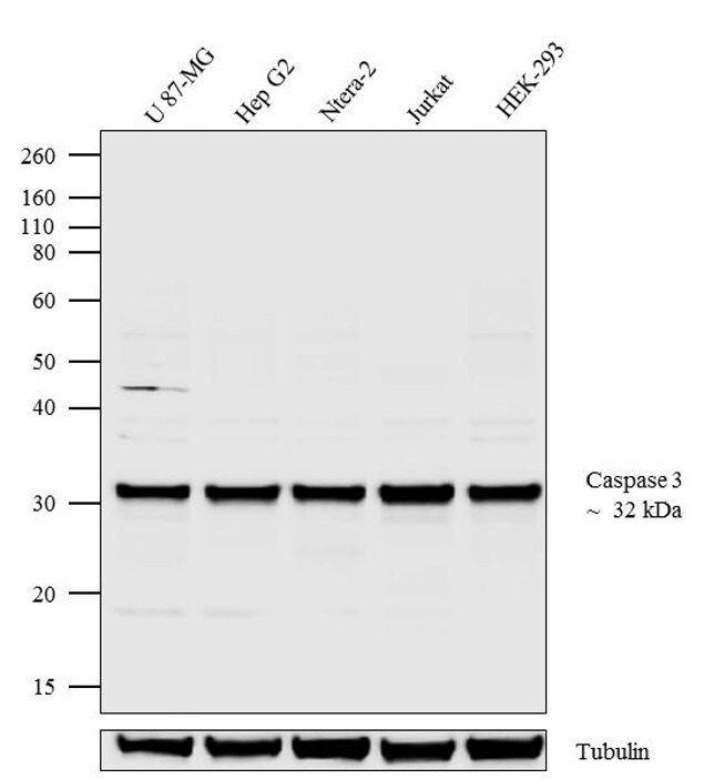
- Western blot analysis of Caspase-3 was performed by loading 20 µg of U-87 MG (lane1), Hep G2 (lane2), Ntera-2 (lane3), Jurkat (lane4) and HEK-293 (lane5) cell lysate using Novex® NuPAGE® 4-12 % Bis-Tris gel (Product # NP0322BOX), XCell SureLock™ Electrophoresis System (Product # EI0002), Novex® Sharp Pre-Stained Protein Standard (LC5800), and iBlot® Dry Blotting System (IB21001). Proteins were transferred to a nitrocellulose membrane and blocked with 5 % skim milk for 1 hour at room temperature. Caspase-3 was detected at ~ 32 kDa using Caspase-3 Mouse Monoclonal Antibody (Product # 43-7800) at 2mg/mL in 5 % skim milk at 4°C overnight on a rocking platform. Goat Anti-Mouse - HRP Secondary Antibody (Product # 62-6520) at 1:4000 dilution was used and chemiluminescent detection was performed using Pierce™ ECL Western Blotting Substrate (Product # 32106).
Supportive validation
- Submitted by
- Invitrogen Antibodies (provider)
- Main image

- Experimental details
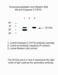
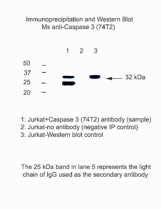
- Immunoprecipitation and Western blot analysis of Caspase 3 using a monoclonal antibody (Product # 43-7800).

Supportive validation
- Submitted by
- Invitrogen Antibodies (provider)
- Main image
- Experimental details
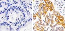
- Immunohistochemistry analysis of Caspase-3 showing staining in the cytoplasm of paraffin-embedded human lung adenocarcinoma tissue (right) compared to a negative control without primary antibody (left). To expose target proteins, antigen retrieval was performed using 10mM sodium citrate (pH 6.0), microwaved for 8-15 min. Following antigen retrieval, tissues were blocked in 3% H2O2-methanol for 15 min at room temperature, washed with ddH2O and PBS, and then probed with a Caspase-3 monoclonal antibody (Product # 43-7800) diluted in 3% BSA-PBS at a dilution of 1:100 overnight at 4ºC in a humidified chamber. Tissues were washed extensively in PBST and detection was performed using an HRP-conjugated secondary antibody followed by colorimetric detection using a DAB kit. Tissues were counterstained with hematoxylin and dehydrated with ethanol and xylene to prep for mounting.

- Submitted by
- Invitrogen Antibodies (provider)
- Main image
- Experimental details
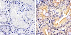
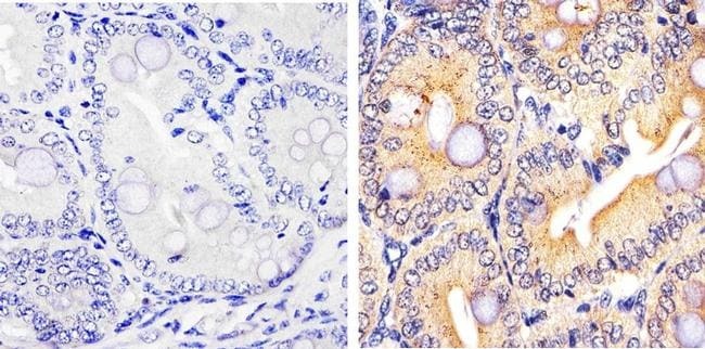
- Immunohistochemistry analysis of Caspase-3 showing staining in the cytoplasm of paraffin-embedded mouse colon tissue (right) compared to a negative control without primary antibody (left). To expose target proteins, antigen retrieval was performed using 10mM sodium citrate (pH 6.0), microwaved for 8-15 min. Following antigen retrieval, tissues were blocked in 3% H2O2-methanol for 15 min at room temperature, washed with ddH2O and PBS, and then probed with a Caspase-3 monoclonal antibody (Product # 43-7800) diluted in 3% BSA-PBS at a dilution of 1:20 overnight at 4ºC in a humidified chamber. Tissues were washed extensively in PBST and detection was performed using an HRP-conjugated secondary antibody followed by colorimetric detection using a DAB kit. Tissues were counterstained with hematoxylin and dehydrated with ethanol and xylene to prep for mounting.
Supportive validation
- Submitted by
- Invitrogen Antibodies (provider)
- Main image
- Experimental details
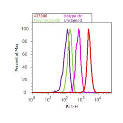
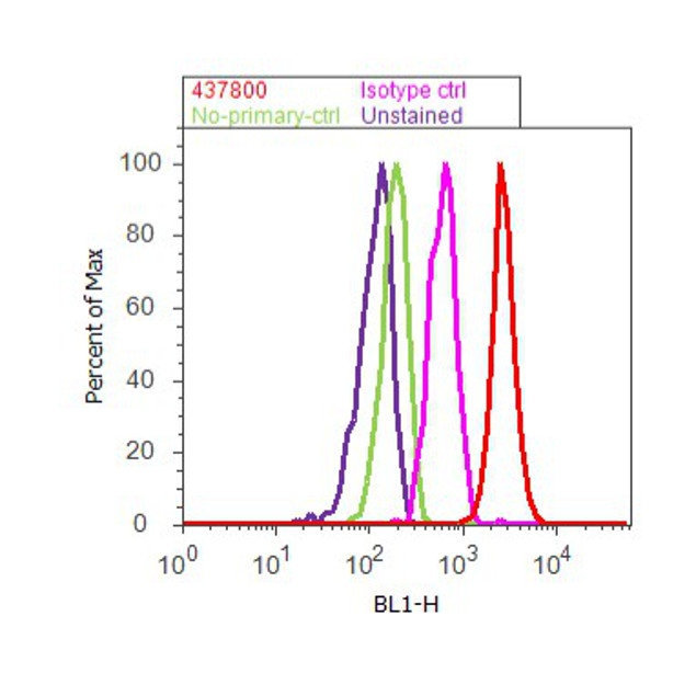
- Flow cytometry analysis of Caspase 3 was done on K562 cells. Cells were fixed with 70% ethanol for 10 minutes, permeabilized with 0.25% Triton™ X-100 for 20 minutes, and blocked with 5% BSA for 30 minutes at room temperature. Cells were labeled with Caspase 3 Mouse Monoclonal Antibody (437800, red histogram) or with mouse isotype control (pink histogram) at 3-5 ug/million cells in 2.5% BSA. After incubation at room temperature for 2 hours, the cells were labeled with Alexa Fluor® 488 Rabbit Anti-Mouse Secondary Antibody (A11059) at a dilution of 1:400 for 30 minutes at room temperature. The representative 10,000 cells were acquired and analyzed for each sample using an Attune® Acoustic Focusing Cytometer. The purple histogram represents unstained control cells and the green histogram represents no-primary-antibody control.
Supportive validation
- Submitted by
- Invitrogen Antibodies (provider)
- Main image
- Experimental details
- Immunoprecipitation and Western blot analysis of Caspase 3 using a monoclonal antibody (Product # 43-7800).
- Submitted by
- Invitrogen Antibodies (provider)
- Main image
- Experimental details
- NULL
- Submitted by
- Invitrogen Antibodies (provider)
- Main image
- Experimental details
- Figure 2. DZX regulates the expression of CHOP, GRP78, caspase-12 and caspase-3 in OGD-treated hippocampal cells. (A) Immunohistochemical staining demonstrated the distribution of GRP78 and CHOP in hippocampal cells exposed to OGD. (B) Western blot analysis was used to detect the expression of GRP78 and CHOP in hippocampal cells exposed to OGD. (C) Protein expression levels of caspase-12 and caspase-3 in hippocampal cells exposed to OGD were detected using western blotting. Data are expressed as the mean +- standard deviation. *P

- Submitted by
- Invitrogen Antibodies (provider)
- Main image

- Experimental details
- Figure 5. Pretreatment with DZX modulates the protein expression of Bcl-2, caspase-12, caspase-3, Bax, CHOP and GRP78 in rats subjected to I/R. Western blot analysis was used to detect the protein expression levels of Bcl-2, caspase-12, caspase-3, Bax, CHOP and GRP78 in hippocampal cells isolated from I/R-treated rats. Rats were treated with DZX with or without 5-HD and subjected to cerebral ischemia for 2 h followed by 12 h of reperfusion. Sham rats were not subjected to cerebral ischemia. DZX, diazoxide; Bcl-2, B-cell lymphoma-2; Bax, Bcl-2-associated X protein; CHOP, C/EBP homologous protein; GRP78, 78 kDa glucose-regulated protein; I/R, ischemia/reperfusion; 5-HD, 5-hydroxydecanoate.
- Submitted by
- Invitrogen Antibodies (provider)
- Main image
- Experimental details
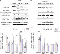
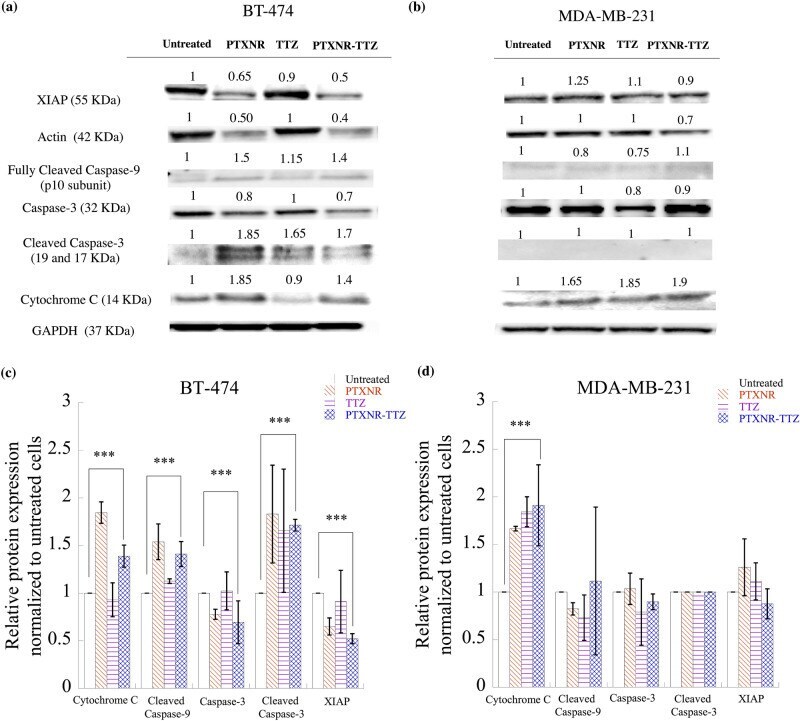
- Figure 7 Western blot analysis: The cell signaling protein expressions of ( a ) BT-474 and ( b ) MDA-MB-231 cells using PBS, PTX alone, TTZ alone, and PTXNR-TTX treatments. The relative intensity of XIAP, actin, cleaved caspase-9, caspase 3, cleaved caspase 3, and cytochrome C is analyzed after 48 h of treatment in ( c ) BT-474 and ( d ) MDA-MB-231 cells using ImageJ/ Fiji. For each protein expression the experiment was replicated at least n = 2-4 times and the average data is presented as mean +- standard deviation. The relative protein expressions are normalized to the untreated control cells and to the housekeeping GAPDH protein expression (hypothesized expression mean, u = 1). The p values for overexpression or downregulation of cytochrome-C, cleaved caspase-3, and XIAP in BT-474 are 0.039, 0.003, and 0.000049, respectively. The p value for the overexpression of cytochrome-C in MDA-MB-231 is 0.047. ***Represents p < 0.05 (for detailed p value calculations, please see SI Table 6 ).
- Submitted by
- Invitrogen Antibodies (provider)
- Main image
- Experimental details
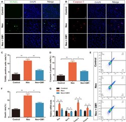
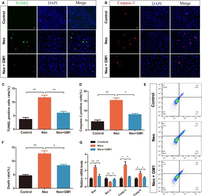
- FIGURE 2 GM1 reduced neomycin-induced apoptosis in HEI-OC-1 cells. (A) Immunofluorescence images of apoptotic HEI-OC-1 cells with TUNEL and DAPI double staining. (B) Immunofluorescence images of apoptotic HEI-OC-1 cells with cleaved CASPASE-3 and DAPI double staining. (C,D) Quantification of the number of apoptotic cells in (A,B) . (E,F) Survival and percentage of HEI-OC-1 cells determined by DAPI/PI staining flow cytometry. (G) Expression of proapoptotic and antiapoptotic genes in HEI-OC-1 cells treated with neomycin and/or GM1 determined by qRT-PCR; the values were normalized to Gapdh as an internal control. The data are expressed as the mean +- SD of triplicate samples. * p < 0.05, ** p < 0.01. Scale bars = 20 mum.
- Submitted by
- Invitrogen Antibodies (provider)
- Main image

- Experimental details
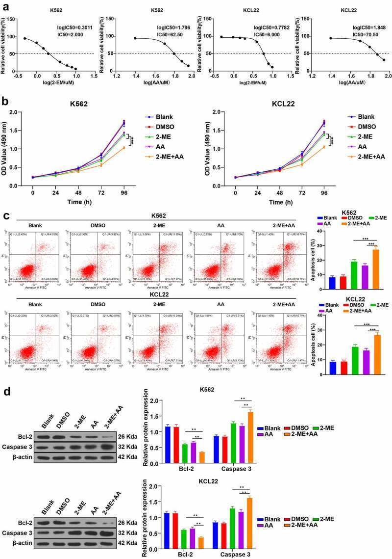
- Figure 1. 2-ME combined with AA promotes CML cell apoptosis. K562 and KCL22 cells were treated with 2-ME and AA. (a) Viability of cells treated with different concentrations of 2-ME and AA for 48 h was measured using MTT assay, and IC50 was calculated. IC50 doses of 2-ME and AA were used for subsequent cell treatment. (b) Cell proliferation activity at different time points examined using MTT assay. (c) Cell apoptosis measured using flow cytometry. (d) Levels of Bcl-2 and Caspase 3 measured using Western blotting. Cell experiment was conducted 3 times independently. Data were described as mean +- standard deviation. Data were analyzed using one-way ANOVA, followed by Tukey's multiple comparison test, ** p < 0.01 vs . DMSO group.
- Submitted by
- Invitrogen Antibodies (provider)
- Main image

- Experimental details
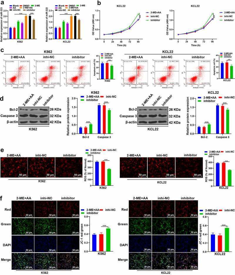
- Figure 3. 2-ME combined with AA upregulates intracellular miR-223 expression. 2-ME + AA treated K562 and KCL22 cells were delivered with miR-223 inhibitor, with inhibitor NC as control. (a) miR-223 expression tested using RT-qPCR. (b) Cell proliferation at different times measured using MTT assay. (c) Cell apoptosis measured using flow cytometry. (d) Protein levels of Bcl-2 and Caspase 3 measured using Western blotting. (e) Content of ROS. F: Change of MMP. Cell experiment was conducted 3 times independently. Data were described as mean +- standard deviation. Data were analyzed using one-way ANOVA, followed by Tukey's multiple comparison test, *** p < 0.001. inhi-NC: 2-ME + AA + inhibitor NC; inhibitor: 2-ME + AA + miR-223 inhibitor.
- Submitted by
- Invitrogen Antibodies (provider)
- Main image
- Experimental details
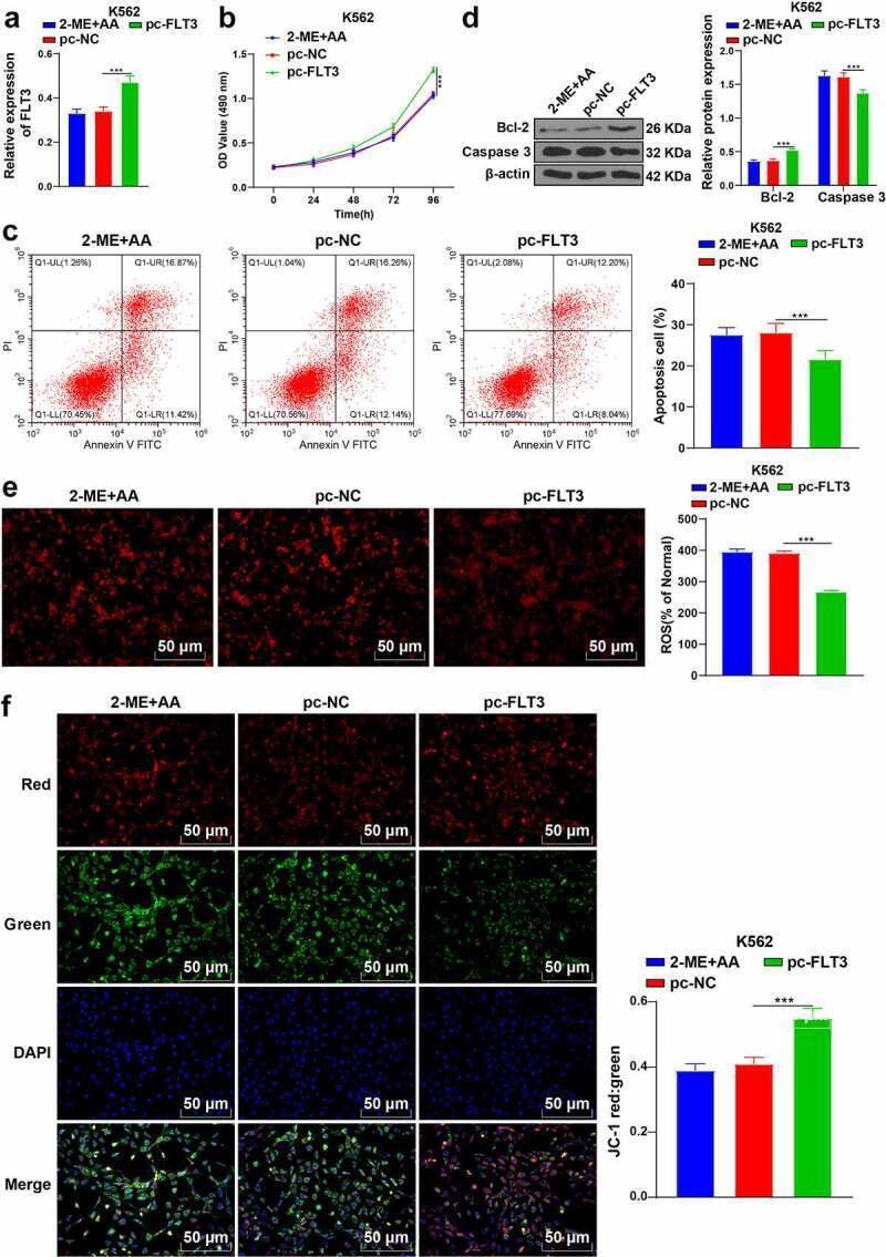
- Figure 5. Upregulation of FLT3 partially reverses the promoting effect of 2-ME + AA on apoptosis of K562 cells. 2-ME + AA treated K562 cells were delivered with pcDNA3.1-FLT3, with pcDNA3.1-NC as control. (a) FLT3 expression tested using RT-qPCR. (b) Cell proliferation at different times measured using MTT assay. (c) Cell apoptosis measured using flow cytometry. (d) Protein levels of Bcl-2 and Caspase 3 examined using Western blotting. (e) Content of ROS. (f) Change of MMP. Cell experiment was conducted 3 times independently. Data were described as mean +- standard deviation. Data were analyzed using one-way ANOVA, followed by Tukey's multiple comparison test, *** p < 0.001. pc-NC: 2-ME + AA + pcDNA3.1-NC; pc-FLT3: 2-ME + AA + pcDNA3.1-FLT3.
- Submitted by
- Invitrogen Antibodies (provider)
- Main image

- Experimental details
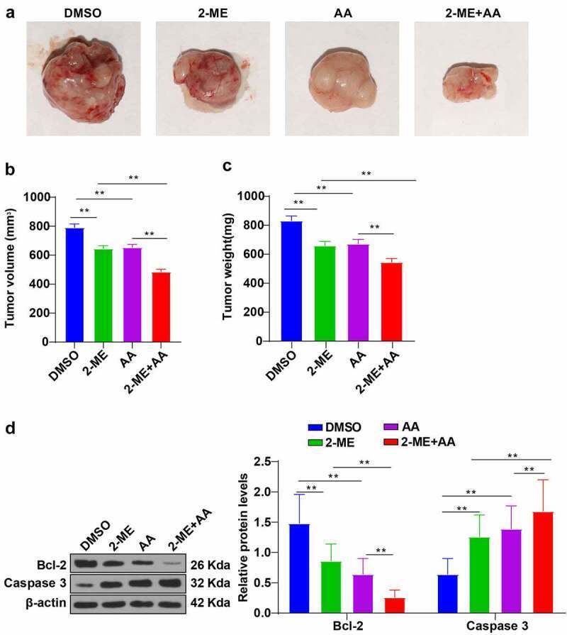
- Figure 7. 2-ME combined with AA inhibited CML xenograft growth in mice. CML xenograft nude mouse models were established, and then intravenously injected with 2-ME, AA alone, or a combination of 2-ME and AA. (a) Image of mouse tumors. (b) Average tumor volume. (c) Average tumor weight. (d) Protein levels of Bcl-2 and Caspase 3 examined using Western blotting. N = 8, data were presented as mean +- standard deviation. Data among multiple groups were analyzed using one-way ANOVA, followed by Tukey's multiple comparison test, ** p < 0.01.
- Submitted by
- Invitrogen Antibodies (provider)
- Main image
- Experimental details
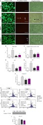
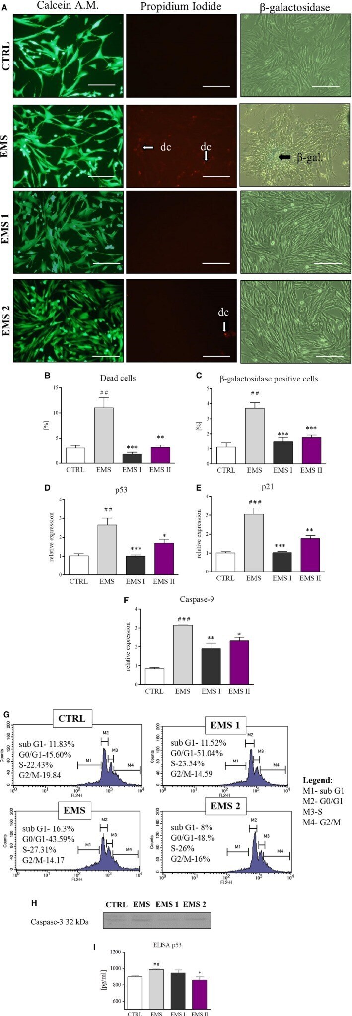
- Figure 3 AZA / RES reversed aging and reduced apoptosis in ASC EMS . In order to evaluate apoptosis in culture cells were cultured for 24 hours in control or experimental ( AZA / RES ) condition. Next, cells were subjected to staining procedures and RT - PCR analysis. In order to visualize live and dead cells in culture Calcein A.M and propidium iodide staining was applied (A). Moreover, senescent cells in cultures were visualized by beta-galactosidase staining (A). Furthermore, data obtained from representative photographs was quantified (B and C). Presented results displayed that AZA / RES treatment markedly reduced number of dead and senescent cells. Apoptosis incidence was also investigated with RT - PCR for p53 (D), p21 (E), and Caspase-9 (F). Representative graphs from cell cycle analysis (G). Western blot for caspase-3 (H) and ELISA for p53 (I), Magnification x100, scale bars: 250 mum. Results expressed as mean +- SD . Statistical significance indicated as asterisk (*) when comparing the result to ASC CTRL , and as hashtag (#) when comparing to ASC EMS . * P < 0.05; ##, ** P < 0.01; ###, *** P < 0.001
- Submitted by
- Invitrogen Antibodies (provider)
- Main image
- Experimental details
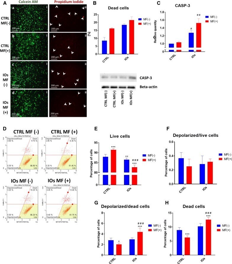
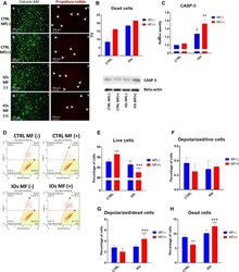
- Fig. 17 IOs and MF affect mitochondrial membrane potential and contributes to enhance apoptosis in osteoclasts. Representative photographs from Calcein A.M. and Propidium iodide ( A ) and their quantification ( B ) revealed increased number of apoptotic cells in IOs and MF groups. Western blot analysis revealed increased amount of CASP-3 in IOs MF- and IOs MF+ groups. To investigate mitochondrial condition in cells Muse (r) MitoPotential analysis was performed ( D ). Based on the obtained data, percentage of live cells ( E ), depolarized/live cells ( F ), depolarized/dead cells ( G ) and dead cells ( H ) was calculated. Results expressed as mean +- SD. Statistical significance indicated as asterisk (*) when comparing the results between corresponding bars representing MF- and MF+ groups, and as number sign (#) when comparing to CTRL MF-. #,*p < 0.05, ##, **p < 0.01, ***, ###p < 0.001