 Explore
 Validate
 Learn
LearnAMAb90988
antibody from Atlas Antibodies
Targeting: PDIA3
ERp57, ERp60, ERp61, GRP57, GRP58, HsT17083, P58, PI-PLC
 Western blot
 Immunocytochemistry
Antibody data
- Antibody Data
- Antigen structure
- References [0]
- Comments [0]
- Validations
- Western blot [3]
- Immunocytochemistry [5]
- Immunohistochemistry [10]
Submit
Validation data
Reference
Comment
Report error
- Product number
- AMAb90988 - Provider product page

- Provider
- Atlas Antibodies
- Proper citation
- Atlas Antibodies Cat#AMAb90988, RRID:AB_2665750
- Product name
- Anti-PDIA3
- Antibody type
- Monoclonal
- Reactivity
- Human
- Host
- Mouse
- Conjugate
- Unconjugated
- Antigen sequence
PTLKIFRDGEEAGAYDGPRTADGIVSHLKKQAGPA
SVPLRTEEEFKKFISDKDASIVGFFDDSFSEAHSE
FLKAASNLRDNYRFAHTNVESLVNEYDDNGEGIIL
FRPSHLTNKFEDK- Epitope
- Binds to an epitope located within the peptide sequence KKFISDKDASIVGFF as determined by overlapping synthetic peptides.
- Isotype
- IgG
- Antibody clone number
- CL2444
- Vial size
- 100 µl
- Storage
- Store at +4°C for short term storage. Long time storage is recommended at -20°C.
No comments: Submit comment
Enhanced validation
- Submitted by
- Atlas Antibodies (provider)
- Enhanced method
- Genetic validation
- Main image

- Experimental details
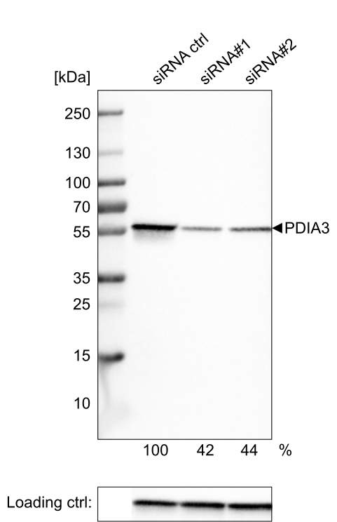
- Western blot analysis in U-251MG cells transfected with control siRNA, target specific siRNA probe #1 and #2, using Anti-PDIA3 antibody. Remaining relative intensity is presented. Loading control: Anti-GAPDH.

- Submitted by
- Atlas Antibodies (provider)
- Main image

- Experimental details
- Lane 1: Marker [kDa]Lane 2:Human cell line U-251 MG

- Submitted by
- Atlas Antibodies (provider)
- Main image

- Experimental details
- Western blot analysis of extracts from U-251 cells, transfected with: control siRNA, target specific siRNA probe #1, target specific siRNA probe #2, using Anti-PDIA3 monoclonal antibody. Downregulation of antibody signal confirms target specificity. Remaining % intensity, relative control lane, is indicated. Anti-GAPDH monoclonal antibody was used as loading control.
Supportive validation
- Submitted by
- Atlas Antibodies (provider)
- Main image

- Experimental details
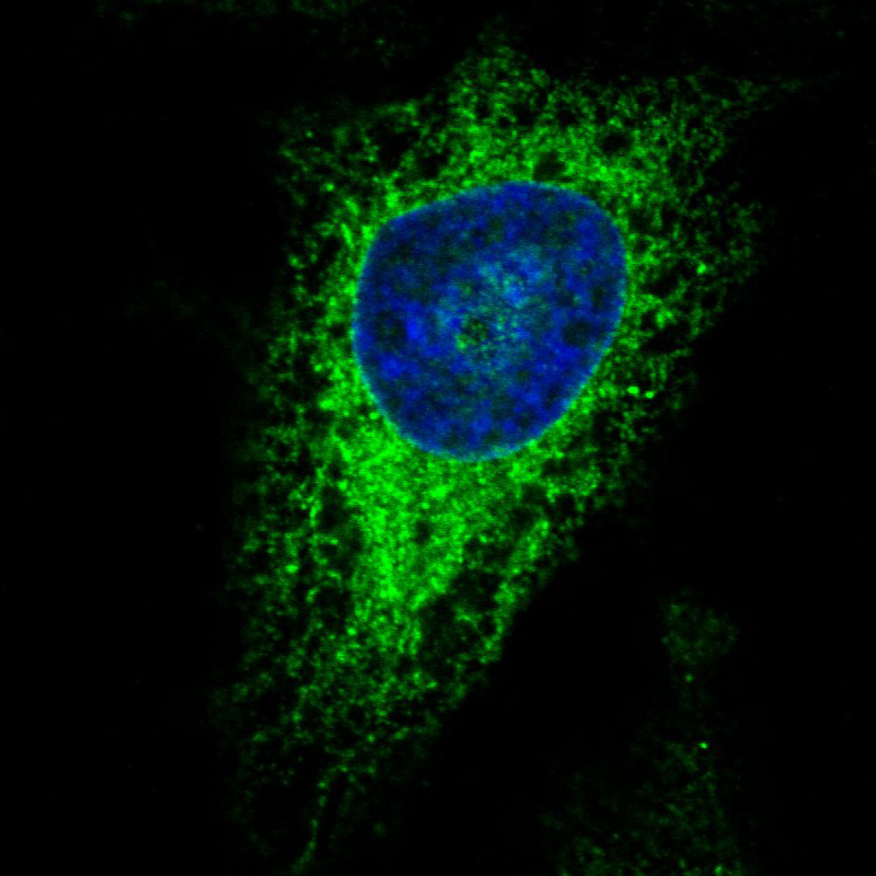
- Immunofluorescence staining in HeLa cell line with Anti-PDIA3 monoclonal antibody, showing specific staining of endoplasmic reticulum in green. Microtubule- and nuclear probes are visualized in red and blue respectively (where available).
- Sample type
- HUMAN
- Submitted by
- Atlas Antibodies (provider)
- Main image

- Experimental details
- Immunofluorescence staining in A431 cell line with Anti-PDIA3 monoclonal antibody, showing specific staining of endoplasmic reticulum in green. Microtubule- and nuclear probes are visualized in red and blue respectively (where available).
- Sample type
- HUMAN

- Submitted by
- Atlas Antibodies (provider)
- Main image

- Experimental details
- Immunofluorescence staining in MCF7 cell line with Anti-PDIA3 monoclonal antibody, showing specific staining of endoplasmic reticulum in green. Microtubule- and nuclear probes are visualized in red and blue respectively (where available).
- Sample type
- HUMAN

- Submitted by
- Atlas Antibodies (provider)
- Main image

- Experimental details
- Immunofluorescence staining in U2OS cell line with Anti-PDIA3 monoclonal antibody, showing specific staining of endoplasmic reticulum in green. Microtubule- and nuclear probes are visualized in red and blue respectively (where available).
- Sample type
- HUMAN

- Submitted by
- Atlas Antibodies (provider)
- Main image

- Experimental details
- Immunofluorescence staining in U251 cell line with Anti-PDIA3 monoclonal antibody, showing specific staining of endoplasmic reticulum in green. Microtubule- and nuclear probes are visualized in red and blue respectively (where available).
- Sample type
- HUMAN

Enhanced validation
Supportive validation
- Submitted by
- Atlas Antibodies (provider)
- Enhanced method
- Orthogonal validation
- Main image
- Experimental details
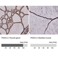
- Immunohistochemistry analysis in human thyroid gland and skeletal muscle tissues using AMAb90988 antibody. Corresponding PDIA3 RNA-seq data are presented for the same tissues.
- Sample type
- HUMAN

Supportive validation
- Submitted by
- Atlas Antibodies (provider)
- Main image

- Experimental details
- Immunohistochemical staining of human cerebral cortex shows strong cytoplasmic immunoreactivity in neurons.

- Submitted by
- Atlas Antibodies (provider)
- Main image

- Experimental details
- Immunohistochemical staining of human prostate shows strong cytoplasmic immunoreactivity in glandular cells.

- Submitted by
- Atlas Antibodies (provider)
- Main image

- Experimental details
- Immunohistochemical staining of human liver shows strong cytoplasmic positivity.

- Submitted by
- Atlas Antibodies (provider)
- Main image

- Experimental details
- Immunohistochemical staining of human rectum shows strong cytoplasmic immunoreactivity in glandular epithelium and lamina propria cells.

- Submitted by
- Atlas Antibodies (provider)
- Main image

- Experimental details
- Immunohistochemical staining of human cerebellum shows strong cytoplasmic positivity in Purkinje cells and molecular layer neurons.

- Submitted by
- Atlas Antibodies (provider)
- Main image

- Experimental details
- Immunohistochemical staining of human thyroid gland shows moderate to strong cytoplasmic positivity in glandular cells.

- Submitted by
- Atlas Antibodies (provider)
- Main image

- Experimental details
- Immunohistochemical staining of human cerebral cortex shows moderate cytoplasmic positivity in neurons.

- Submitted by
- Atlas Antibodies (provider)
- Main image

- Experimental details
- Immunohistochemical staining of human endometrium shows moderate cytoplasmic positivity in glandular cells.

- Submitted by
- Atlas Antibodies (provider)
- Main image

- Experimental details
- Immunohistochemical staining of human skeletal muscle shows no positivity in striated muscle fibers as expected.
