 Explore
 Validate
 Learn
LearnNBP1-84797
antibody from Novus Biologicals
Targeting: PDIA3
ERp57, ERp60, ERp61, GRP57, GRP58, HsT17083, P58, PI-PLC
 Western blot
 Immunocytochemistry
Antibody data
- Antibody Data
- Antigen structure
- References [3]
- Comments [0]
- Validations
- Western blot [2]
- Immunohistochemistry [10]
Submit
Validation data
Reference
Comment
Report error
- Product number
- NBP1-84797 - Provider product page

- Provider
- Novus Biologicals
- Proper citation
- Novus Cat#NBP1-84797, RRID:AB_11027170
- Product name
- Rabbit Polyclonal ERp57/PDIA3 Antibody
- Antibody type
- Polyclonal
- Description
- Immunogen affinity purified. Specificity of human ERp57/PDIA3 antibody verified on a Protein Array containing target protein plus 383 other non-specific proteins.
- Reactivity
- Human
- Host
- Rabbit
- Isotype
- IgG
- Vial size
- 0.1 ml
- Storage
- Store at 4C short term. Aliquot and store at -20C long term. Avoid freeze-thaw cycles.
Submitted references The dehydrogenase region of the NADPH oxidase component Nox2 acts as a protein disulfide isomerase (PDI) resembling PDIA3 with a role in the binding of the activator protein p67 (phox.).
SILAC-based quantitative proteomic approach to identify potential biomarkers from the esophageal squamous cell carcinoma secretome.
SILAC-based quantitative proteomic approach to identify potential biomarkers from the esophageal squamous cell carcinoma secretome.
Bechor E, Dahan I, Fradin T, Berdichevsky Y, Zahavi A, Federman Gross A, Rafalowski M, Pick E
Frontiers in chemistry 2015;3:3
Frontiers in chemistry 2015;3:3
SILAC-based quantitative proteomic approach to identify potential biomarkers from the esophageal squamous cell carcinoma secretome.
Kashyap MK, Harsha HC, Renuse S, Pawar H, Sahasrabuddhe NA, Kim MS, Marimuthu A, Keerthikumar S, Muthusamy B, Kandasamy K, Subbannayya Y, Prasad TS, Mahmood R, Chaerkady R, Meltzer SJ, Kumar RV, Rustgi AK, Pandey A
Cancer biology & therapy 2010 Oct 15;10(8):796-810
Cancer biology & therapy 2010 Oct 15;10(8):796-810
No comments: Submit comment
Supportive validation
- Submitted by
- Novus Biologicals (provider)
- Main image
- Experimental details
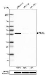
- Western Blot: ERp57/PDIA3 Antibody [NBP1-84797] - Analysis in U-251MG cells transfected with control siRNA, target specific siRNA probe #1 and #2. Remaining relative intensity is presented. Loading control: Anti-GAPDH.

- Submitted by
- Novus Biologicals (provider)
- Main image
- Experimental details
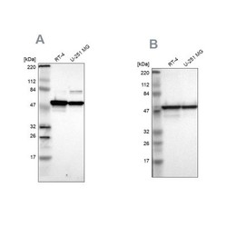
- Western Blot: ERp57/PDIA3 Antibody [NBP1-84797] - Analysis using Anti-PDIA3 antibody NBP1-84797 (A) shows similar pattern to independent antibody NBP1-84796 (B).

Supportive validation
- Submitted by
- Novus Biologicals (provider)
- Main image
- Experimental details
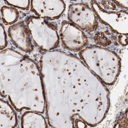
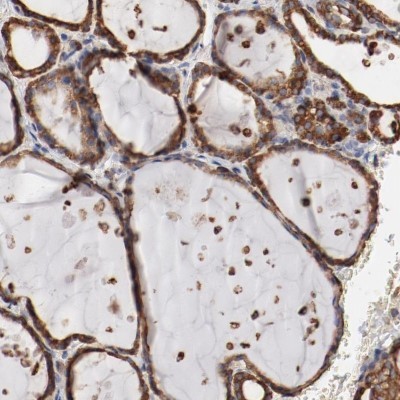
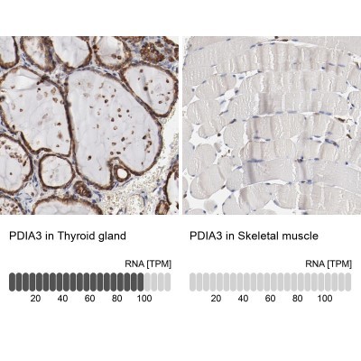
- Immunohistochemistry-Paraffin: ERp57/PDIA3 Antibody [NBP1-84797] - Staining of human thyroid gland shows high expression.
- Submitted by
- Novus Biologicals (provider)
- Main image
- Experimental details
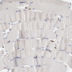
- Immunohistochemistry-Paraffin: ERp57/PDIA3 Antibody [NBP1-84797] - Staining of human skeletal muscle shows low expression as expected.
- Submitted by
- Novus Biologicals (provider)
- Main image

- Experimental details
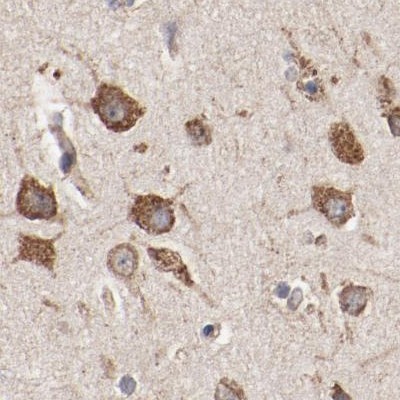
- Immunohistochemistry-Paraffin: ERp57/PDIA3 Antibody [NBP1-84797] - Staining of human cerebral cortex.
- Submitted by
- Novus Biologicals (provider)
- Main image

- Experimental details
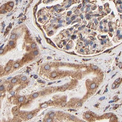
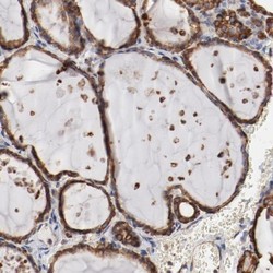
- Immunohistochemistry-Paraffin: ERp57/PDIA3 Antibody [NBP1-84797] - Staining of human kidney.
- Submitted by
- Novus Biologicals (provider)
- Main image
- Experimental details
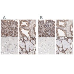
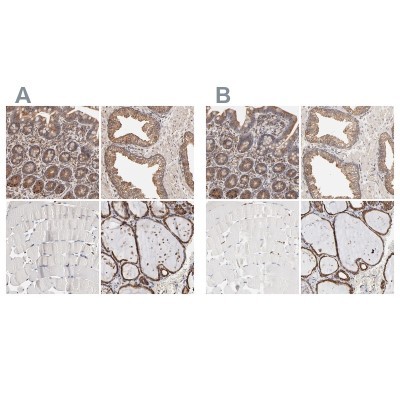
- Immunohistochemistry-Paraffin: ERp57/PDIA3 Antibody [NBP1-84797] - Staining of human gastrointestinal, prostate, skeletal muscle and thyroid gland using Anti-PDIA3 antibody NBP1-84797 (A) shows similar protein distribution across tissues to independent antibody NBP1-84796 (B).
- Submitted by
- Novus Biologicals (provider)
- Main image

- Experimental details
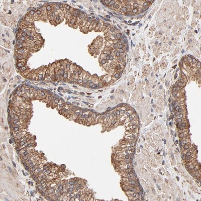
- Immunohistochemistry-Paraffin: ERp57/PDIA3 Antibody [NBP1-84797] - Staining of human prostate shows moderate cytoplasmic positivity in glandular cells.
- Submitted by
- Novus Biologicals (provider)
- Main image
- Experimental details
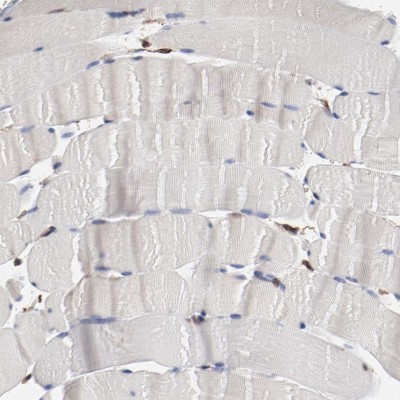
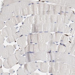
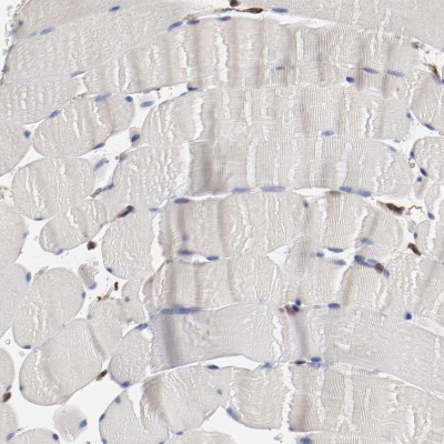
- Immunohistochemistry-Paraffin: ERp57/PDIA3 Antibody [NBP1-84797] - Staining of human skeletal muscle shows no cytoplasmic positivity in myocytes as expected.
- Submitted by
- Novus Biologicals (provider)
- Main image
- Experimental details
- Immunohistochemistry-Paraffin: ERp57/PDIA3 Antibody [NBP1-84797] - Staining of human thyroid gland shows strong cytoplasmic positivity in glandular cells.

- Submitted by
- Novus Biologicals (provider)
- Main image
- Experimental details
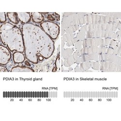
- Immunohistochemistry-Paraffin: ERp57/PDIA3 Antibody [NBP1-84797] - Staining in human thyroid gland and skeletal muscle tissues using NBP1-84797 antibody. Corresponding PDIA3 RNA-seq data are presented for the same tissues.
- Submitted by
- Novus Biologicals (provider)
- Main image
- Experimental details
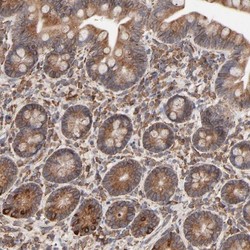
- Immunohistochemistry-Paraffin: ERp57/PDIA3 Antibody [NBP1-84797] - Staining of human small intestine shows moderate to strong cytoplasmic positivity in glandular cells.
