 Explore
 Validate
 Learn
Learn Western blot
 Immunocytochemistry
Antibody data
- Antibody Data
- Antigen structure
- References [5]
- Comments [0]
- Validations
- Western blot [3]
- Immunohistochemistry [2]
- Flow cytometry [1]
Submit
Validation data
Reference
Comment
Report error
- Product number
- NB100-687 - Provider product page

- Provider
- Novus Biologicals
- Proper citation
- Novus Cat#NB100-687, RRID:AB_2265512
- Product name
- Rabbit Polyclonal Cytokeratin 19 Antibody
- Antibody type
- Polyclonal
- Description
- Immunogen affinity purified.
- Reactivity
- Human, Mouse, Rat
- Host
- Rabbit
- Isotype
- IgG
- Vial size
- 0.1 ml
- Concentration
- 1.08 mg/ml
- Storage
- Store at 4C short term. Aliquot and store at -20C long term. Avoid freeze-thaw cycles.
Submitted references Single-cell RNA sequencing demonstrates the molecular and cellular reprogramming of metastatic lung adenocarcinoma.
Impaired liver regeneration in aged mice can be rescued by silencing Hippo core kinases MST1 and MST2.
Bile acids induce hepatic differentiation of mesenchymal stem cells.
Hepatic stellate cells contribute to progenitor cells and liver regeneration.
Death receptor 5 mediated-apoptosis contributes to cholestatic liver disease.
Kim N, Kim HK, Lee K, Hong Y, Cho JH, Choi JW, Lee JI, Suh YL, Ku BM, Eum HH, Choi S, Choi YL, Joung JG, Park WY, Jung HA, Sun JM, Lee SH, Ahn JS, Park K, Ahn MJ, Lee HO
Nature communications 2020 May 8;11(1):2285
Nature communications 2020 May 8;11(1):2285
Impaired liver regeneration in aged mice can be rescued by silencing Hippo core kinases MST1 and MST2.
Loforese G, Malinka T, Keogh A, Baier F, Simillion C, Montani M, Halazonetis TD, Candinas D, Stroka D
EMBO molecular medicine 2017 Jan;9(1):46-60
EMBO molecular medicine 2017 Jan;9(1):46-60
Bile acids induce hepatic differentiation of mesenchymal stem cells.
Sawitza I, Kordes C, Götze S, Herebian D, Häussinger D
Scientific reports 2015 Aug 25;5:13320
Scientific reports 2015 Aug 25;5:13320
Hepatic stellate cells contribute to progenitor cells and liver regeneration.
Kordes C, Sawitza I, Götze S, Herebian D, Häussinger D
The Journal of clinical investigation 2014 Dec;124(12):5503-15
The Journal of clinical investigation 2014 Dec;124(12):5503-15
Death receptor 5 mediated-apoptosis contributes to cholestatic liver disease.
Takeda K, Kojima Y, Ikejima K, Harada K, Yamashina S, Okumura K, Aoyama T, Frese S, Ikeda H, Haynes NM, Cretney E, Yagita H, Sueyoshi N, Sato N, Nakanuma Y, Smyth MJ, Okumura K
Proceedings of the National Academy of Sciences of the United States of America 2008 Aug 5;105(31):10895-900
Proceedings of the National Academy of Sciences of the United States of America 2008 Aug 5;105(31):10895-900
No comments: Submit comment
Supportive validation
- Submitted by
- Novus Biologicals (provider)
- Main image

- Experimental details
- Simple Western: Cytokeratin 19 Antibody [NB100-687] - Lane view shows a specific band for Cytokeratin 19 in 0.5 mg/ml of HepG2 lysate. This experiment was performed under reducing A246 conditions using the 12-230 kDa separation system.

- Submitted by
- Novus Biologicals (provider)
- Main image

- Experimental details
- Western Blot: Cytokeratin 19 Antibody [NB100-687] - Analysis of Cytokeratin 19 in 1) HepG2 and 2) MCF7 lysates.

- Submitted by
- Novus Biologicals (provider)
- Main image

- Experimental details
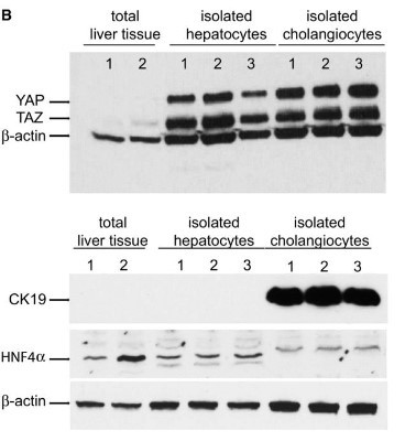
- Western Blot: Cytokeratin 19 Antibody [NB100-687] - Western blot detection of YAP and TAZ in proteins extracted from isolated hepatocytes and cholangiocytes and total liver tissue. Antibodies against CK19 and HNF4a were used to confirm purity of the populations and b-actin was used as a loading control. Representative results from a single experiment with n = 3 independent cell isolations and n = 2 total liver tissues. Image collected and cropped by CiteAb from the following publication(http://embomolmed.embopress.org/lookup/doi/10.15252/emmm.201506089), licensed under a CC-BY licence.
Supportive validation
- Submitted by
- Novus Biologicals (provider)
- Main image

- Experimental details
- Immunohistochemistry: Cytokeratin 19 Antibody [NB100-687] - Staining of Cytokeratin 19 in human kidney carcinoma using DAB with hematoxylin counterstain.

- Submitted by
- Novus Biologicals (provider)
- Main image

- Experimental details
- Immunohistochemistry: Cytokeratin 19 Antibody [NB100-687] - Inhibition of MST improves liver regeneration in aged mice. Representative photomicrograph of an aged liver treated with siMST and stained with H&E and by IHC for CK19 and Ki67. Arrows indicate ductal regions with no signs of reaction or oval cell expansion. Image collected and cropped by CiteAb from the following publication (http://embomolmed.embopress.org/lookup/doi/10.15252/emmm.201506089), licensed under a CC-BY licence.

Supportive validation
- Submitted by
- Novus Biologicals (provider)
- Main image

- Experimental details
- Flow Cytometry: Cytokeratin 19 Antibody [NB100-687] - An intracellular stain was performed on MCF7 cells with Cytokeratin 19 Antibody NB100-687AF647 (blue) and a matched isotype control (orange). Cells were fixed with 4% PFA and then permeabilized with 0.1% saponin. Cells were incubated in an antibody dilution of 2.5 ug/mL for 30 minutes at room temperature. Both antibodies were conjugated to Alexa Fluor 647.
