 Explore
 Validate
 Learn
Learn Western blot
 ELISA
ELISAAntibody data
- Antibody Data
- Antigen structure
- References [0]
- Comments [0]
- Validations
- Western blot [4]
- Flow cytometry [1]
Submit
Validation data
Reference
Comment
Report error
- Product number
- MA1-46375 - Provider product page

- Provider
- Invitrogen Antibodies
- Product name
- TRF1 Monoclonal Antibody (57-6)
- Antibody type
- Monoclonal
- Antigen
- Recombinant full-length protein
- Description
- This antibody will not work in ICC/IF applications. Suggested positive control: HeLa nuclear extracts, antigen standard for TERF1 (transient overexpression lysate), Hela whole cell extract.
- Reactivity
- Human, Mouse, Rat
- Host
- Mouse
- Isotype
- IgG
- Antibody clone number
- 57-6
- Vial size
- 100 µL
- Concentration
- 1 mg/mL
- Storage
- Store at 4°C short term. For long term storage, store at -20°C, avoiding freeze/thaw cycles.
No comments: Submit comment
Supportive validation
- Submitted by
- Invitrogen Antibodies (provider)
- Main image

- Experimental details
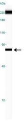
- Western Blot detection of TRF1 in HeLa whole cell extracts

- Submitted by
- Invitrogen Antibodies (provider)
- Main image

- Experimental details
- Western blot analysis of TRF1 in 50 µg lysate. Samples were incubated in TRF1 monoclonal antibody (Product # MA1-46375.

- Submitted by
- Invitrogen Antibodies (provider)
- Main image

- Experimental details
- Western blot analysis of TRF1 in HeLa whole cell extracts. Sample was incubated in TRF1 monoclonal antibody (Product # MA1-46375).

- Submitted by
- Invitrogen Antibodies (provider)
- Main image
- Experimental details
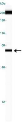
- Western blot analysis of TRF1 in 0.5 mg/mL HeLa lysate. Samples were incubated in TRF1 monoclonal antibody (Product # MA1-46375). This experiment was performed under reducing conditions using the 12-230 kDa separation system..
Supportive validation
- Submitted by
- Invitrogen Antibodies (provider)
- Main image
- Experimental details
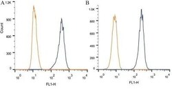
- Flow cytometry of TRF1 in 1 x 10^6 CHO (A) and HEK-293 (B) cells. Samples were incubated in TRF1 monoclonal antibody (Product # MA1-46375) using a dilution of 1 µg/1x10^6 cells. Antibody (dark blue). Isotype control shown in orange.
