 Explore
 Validate
 Learn
Learn Western blot
Antibody data
- Antibody Data
- Antigen structure
- References [0]
- Comments [0]
- Validations
- Western blot [2]
- Immunohistochemistry [1]
Submit
Validation data
Reference
Comment
Report error
- Product number
- ANX-013-200UL - Provider product page

- Provider
- Invitrogen Antibodies
- Product name
- NCX3 (SLC8A3) Polyclonal Antibody
- Antibody type
- Polyclonal
- Antigen
- Other
- Reactivity
- Human, Mouse, Rat
- Host
- Rabbit
- Isotype
- IgG
- Vial size
- 200 µL
- Concentration
- 0.8 mg/mL
- Storage
- -20° C, Avoid Freeze/Thaw Cycles
No comments: Submit comment
Supportive validation
- Submitted by
- Invitrogen Antibodies (provider)
- Main image

- Experimental details
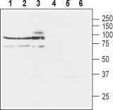
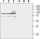
- Western blot analysis of rat brain lysates (lanes 1 and 4), mouse brain lysates (lanes 2 and 5) and human SH-SY5Y neuroblastoma cell lysates (lanes 3 and 6): - 1-3. Anti-NCX3 (SLC8A3) Antibody (#ANX-013), (1:200).4-6. Anti-NCX3 (SLC8A3) Antibody , preincubated with NCX3/SLC8A3 Blocking Peptide (#BLP-NX013).
- Submitted by
- Invitrogen Antibodies (provider)
- Main image

- Experimental details
- Western blot analysis of rat brain lysates (lanes 1 and 4), mouse brain lysates (lanes 2 and 5) and human SH-SY5Y neuroblastoma cell lysates (lanes 3 and 6): - 1-3. Anti-NCX3 (SLC8A3) Antibody (#ANX-013), (1:200).4-6. Anti-NCX3 (SLC8A3) Antibody , preincubated with NCX3/SLC8A3 Blocking Peptide (#BLP-NX013).
Supportive validation
- Submitted by
- Invitrogen Antibodies (provider)
- Main image
- Experimental details
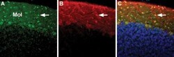
- Expression of NCX3 in rat cerebellum - Immunohistochemical staining of immersion-fixed, free floating rat brain frozen sections using Anti-NCX3 (SLC8A3) Antibody (#ANX-013), (1:100). A. NCX3 staining (green) is expressed mostly in molecular layer (Mol) interneurons (arrow). B. The same section, stained with Parvalbumin (Red). C. Merge of A and B demonstrates that NCX3 appears to be expressed in several GABAergic neurons in the molecular layer of which Parvalbumin positive cells are only one sub-group (see arrow). DAPI counterstain (blue) displays the layout of cerebellar layers.
