Explore
 Validate
 Learn
Learn Immunohistochemistry
Antibody data
- Antibody Data
- Antigen structure
- References [1]
- Comments [0]
- Validations
- Immunohistochemistry [2]
- Other assay [2]
Submit
Validation data
Reference
Comment
Report error
- Product number
- PA5-33529 - Provider product page

- Provider
- Invitrogen Antibodies
- Product name
- PAR3 Polyclonal Antibody
- Antibody type
- Polyclonal
- Antigen
- Synthetic peptide
- Description
- Percent identity with other species by BLAST analysis: Human, Gorilla, Marmoset (100%) Monkey, Bovine, Panda (95%) Gibbon, Dog, Elephant, Pig (89%) Rat, Hamster (84%).
- Reactivity
- Human
- Host
- Rabbit
- Isotype
- IgG
- Vial size
- 50 µg
- Concentration
- 1 mg/mL
- Storage
- Store at 4°C short term. For long term storage, store at -20°C, avoiding freeze/thaw cycles.
Submitted references Contractile forces at tricellular contacts modulate epithelial organization and monolayer integrity.
Salomon J, Gaston C, Magescas J, Duvauchelle B, Canioni D, Sengmanivong L, Mayeux A, Michaux G, Campeotto F, Lemale J, Viala J, Poirier F, Minc N, Schmitz J, Brousse N, Ladoux B, Goulet O, Delacour D
Nature communications 2017 Jan 13;8:13998
Nature communications 2017 Jan 13;8:13998
No comments: Submit comment
Supportive validation
- Submitted by
- Invitrogen Antibodies (provider)
- Main image
- Experimental details
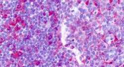
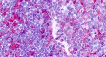
- Immunohistochemical analysis of formalin-fixed paraffin-embedded human tonsil using a F2RL2/PAR3 polyclonal antibody (Product # PA5-33529) at a 2.8-4.6 µg/mL dilution. Heat-induced antigen retrieval was performed prior to staining.
- Submitted by
- Invitrogen Antibodies (provider)
- Main image

- Experimental details
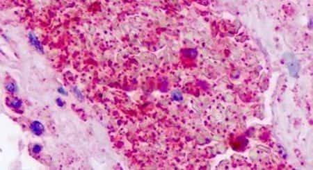
- Immunohistochemical analysis of formalin-fixed paraffin-embedded human platelets using a F2RL2/PAR3 polyclonal antibody (Product # PA5-33529) at a 2.8-4.6 µg/mL dilution. Heat-induced antigen retrieval was performed prior to staining.
Supportive validation
- Submitted by
- Invitrogen Antibodies (provider)
- Main image

- Experimental details
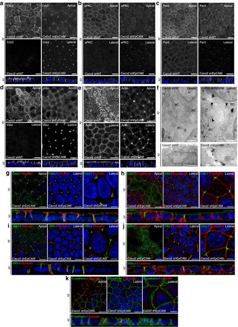
- Figure 3 Apical domain is unusually displaced at tricellular junctions through EpCAM silencing. ( a - e ) Confocal microscopy analysis of Crb3 ( a ), aPKC ( b ), Par3 ( c ), villin ( d ) and actin ( e ) on the apical or lateral sides in control ( Caco2 shNT , left panels) and EpCAM-depleted ( Caco2 shEpCAM , right panels) cells. xy and xz views are presented showing the relocated apical markers at tricellular junctions. Scale bars, 5 mum. ( f ) TEM ultrastructural analysis of tricellular contacts in control ( Caco2 shNT , left) and EpCAM-depleted ( Caco2 shEpCAM , right) cells, showing microvilli at tricellular contacts. Arrowheads point to TJs. Longitudinal ( xy ) and transversal views ( xz ) are presented. BC, bicellular contact, N, nucleus. Scale bars xy , 1 mum and xz , 2 mum. ( g - k ) Confocal microscopy analysis of double immunostainings for Crb3 (green) and occluding (red) ( g ), Crb3 (green) and E-cadherin (red) ( h ), villin (green) and occluding (red) ( i ), villin (green) and E-cadherin (red) ( j ), and E-cadherin (green) and occluding (red) ( k ) on the apical or lateral sides in EpCAM-depleted ( Caco2 shEpCAM ) cells. xy and xz views are presented. Scale bars, 5 mum; high magnifications, 2 mum. Nuclei were detected with Hoechst 33342 staining (blue).
- Submitted by
- Invitrogen Antibodies (provider)
- Main image
- Experimental details
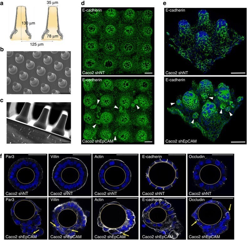
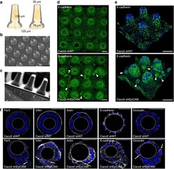
- Figure 8 Generation of PDMS culture inserts that mimic villus topography, and highlighting of tuft-like structures in cultures EpCAM-silenced on synthetic villi. ( a ) Schematic representation of the synthetic polydimethylsiloxane (PDMS) villus inserts. ( b , c ) Scanning electron microscopy analyses of villus PDMS culture inserts: top ( b ) and side ( c ) views are presented. Scale bars, 50 mum ( b ) and 100 mum ( c ). ( d , e ) Confocal microscopy analysis of E-cadherin distribution in control ( Caco2 shNT , upper panels) and EpCAM-silenced ( Caco2 shEpCAM , lower panels) cells that were grown on 3D villus-like micropatterned PDMS inserts for 21 days. After z-stack acquisitions, 3D rendering was generated. White arrowheads point on tuft-like structures. Scale bars, 50 mum. ( f ) Confocal microscopy analysis of Par3, villin, actin, E-cadherin and occludin distribution in control ( Caco2 shNT ) and EpCAM-silenced ( Caco2 shEpCAM ) cells that were grown on villous PDMS inserts for 21 days. Transversal xy views are presented. Par3, villin, actin and occludin display abnormal lateral membrane localization in the absence of EpCAM (yellow arrows). Scale bars, 50 mum.