 Explore
 Validate
 Learn
Learn Western blot
 Immunocytochemistry
Antibody data
- Antibody Data
- Antigen structure
- References [3]
- Comments [0]
- Validations
- Immunocytochemistry [1]
- Immunohistochemistry [4]
Submit
Validation data
Reference
Comment
Report error
- Product number
- HPA003570 - Provider product page

- Provider
- Atlas Antibodies
- Proper citation
- Atlas Antibodies Cat#HPA003570, RRID:AB_1856466
- Product name
- Anti-RPS20
- Antibody type
- Polyclonal
- Reactivity
- Human
- Host
- Rabbit
- Conjugate
- Unconjugated
- Antigen sequence
MAFKDTGKTPVEPEVAIHRIRITLTSRNVKSLEKV
CADLIRGAKEKNLKVKGPVRMPTKTLRITTRKTPC
GEGSKTWDRFQMRIHKRLIDLHSPSEIVKQITSIS
IEPGVEVEVTIA- Isotype
- IgG
- Vial size
- 100 µl
- Storage
- Store at +4°C for short term storage. Long time storage is recommended at -20°C.
Submitted references Translational control of the cytosolic stress response by mitochondrial ribosomal protein L18
Systematic validation of antibody binding and protein subcellular localization using siRNA and confocal microscopy
Posttranscriptional down-regulation of small ribosomal subunit proteins correlates with reduction of 18S rRNA in RPS19 deficiency.
Zhang X, Gao X, Coots R, Conn C, Liu B, Qian S
Nature Structural & Molecular Biology 2015 April
Nature Structural & Molecular Biology 2015 April
Systematic validation of antibody binding and protein subcellular localization using siRNA and confocal microscopy
Stadler C, Hjelmare M, Neumann B, Jonasson K, Pepperkok R, Uhlén M, Lundberg E
Journal of Proteomics 2012 April;75(7):2236-2251
Journal of Proteomics 2012 April;75(7):2236-2251
Posttranscriptional down-regulation of small ribosomal subunit proteins correlates with reduction of 18S rRNA in RPS19 deficiency.
Badhai J, Fröjmark AS, Razzaghian HR, Davey E, Schuster J, Dahl N
FEBS letters 2009 Jun 18;583(12):2049-53
FEBS letters 2009 Jun 18;583(12):2049-53
No comments: Submit comment
Supportive validation
- Submitted by
- Atlas Antibodies (provider)
- Main image

- Experimental details
- Immunofluorescent staining of human cell line U-2 OS shows localization to cytosol & endoplasmic reticulum.
- Sample type
- HUMAN

Supportive validation
- Submitted by
- Atlas Antibodies (provider)
- Main image

- Experimental details
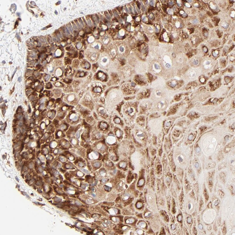
- Immunohistochemical staining of human oral mucosa shows strong cytoplasmic positivity in squamous epithelial cells.
- Submitted by
- Atlas Antibodies (provider)
- Main image

- Experimental details
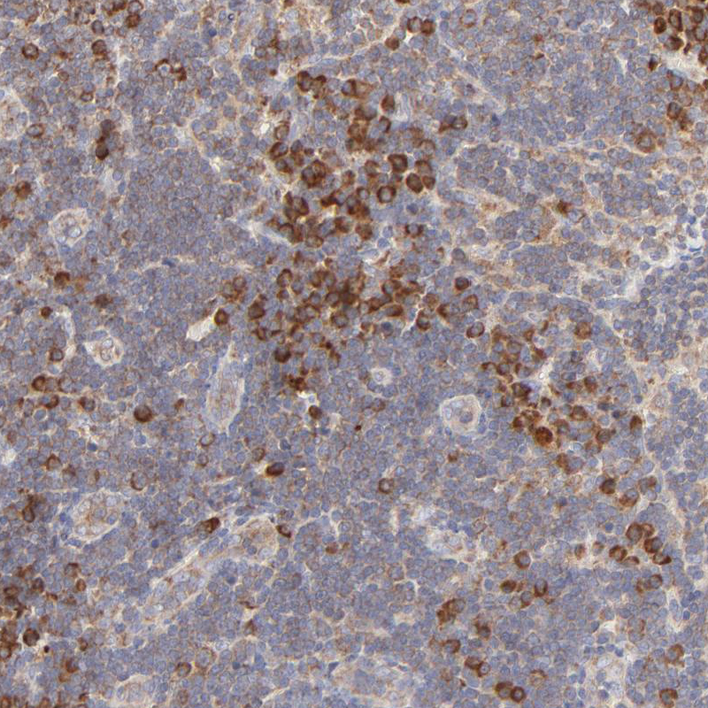
- Immunohistochemical staining of human lymph node shows moderate cytoplasmic positivity in lymphoid cells.
- Sample type
- HUMAN
- Submitted by
- Atlas Antibodies (provider)
- Main image
- Experimental details
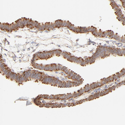
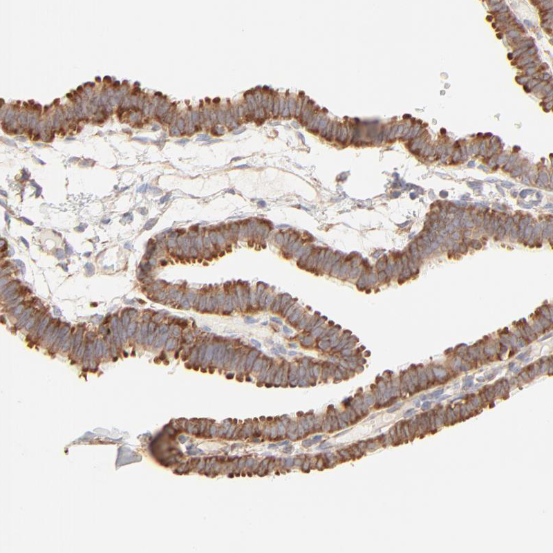
- Immunohistochemical staining of human fallopian tube shows moderate positivity in glandular cells.
- Sample type
- HUMAN
- Submitted by
- Atlas Antibodies (provider)
- Main image
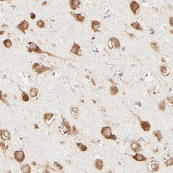
- Experimental details
- Immunohistochemical staining of human cerebral cortex shows moderate cytoplasmic positivity in neuronal cells.
- Sample type
- HUMAN
