Explore
 Validate
 Learn
Learn Western blot
 Immunoprecipitation
Antibody data
- Antibody Data
- Antigen structure
- References [0]
- Comments [0]
- Validations
- Western blot [10]
- Immunocytochemistry [2]
- Immunohistochemistry [6]
- Other assay [1]
Submit
Validation data
Reference
Comment
Report error
- Product number
- 15066-1-AP - Provider product page

- Provider
- Invitrogen Antibodies
- Product name
- NDUFS3 Polyclonal Antibody
- Antibody type
- Polyclonal
- Antigen
- Other
- Description
- Immunogen sequence: MAAAAVARL WWRGILGASA LTRGTGRPSV LLLPVRRESA GADTRPTVRP RNDVAHKQLS AFGEYVAEIL PKYVQQVQVS CFNELEVCIH PDGVIPVLTF LRDHTNAQFK SLVDLTAVDV PTRQNRFEIV YNLLSLRFNS RIRVKTYTDE LTPIESAVSV FKAANWYERE IWDMFGVFFA NHPDLRRILT DYGFEGHPFR KDFPLSGYVE LRYDDEVKRV VAEPVELAQE FRKFDLNSPW EAFPVYRQPP ESLKLEAGDK KPDAK (1-264 aa encoded by BC000617)
- Reactivity
- Human, Mouse, Rat
- Host
- Rabbit
- Isotype
- IgG
- Vial size
- 150 µL
- Concentration
- 0.21 mg/mL
- Storage
- -20°C
No comments: Submit comment
Supportive validation
- Submitted by
- Invitrogen Antibodies (provider)
- Main image
- Experimental details
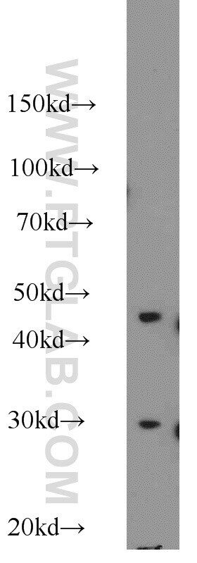
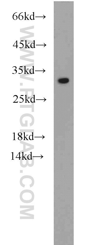
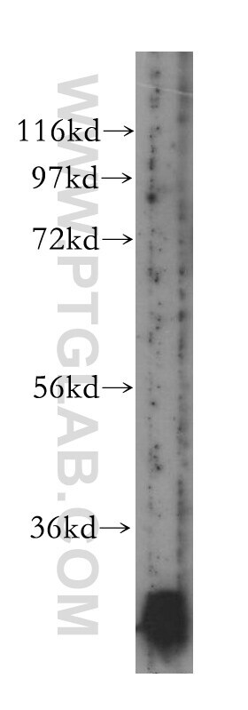
- Mouse heart tissue were subjected to SDS PAGE followed by western blot with 15066-1-AP (NDUFS3 antibody) at dilution of 1:500 incubated at room temperature for 1.5 hours.

- Submitted by
- Invitrogen Antibodies (provider)
- Main image
- Experimental details
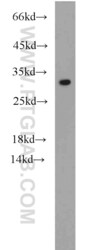
- Human skeletal muscle tissue were subjected to SDS PAGE followed by western blot with 15066-1-AP (NDUFS3 antibody) at dilution of 1:200 incubated at room temperature for 1.5 hours.
- Submitted by
- Invitrogen Antibodies (provider)
- Main image
- Experimental details
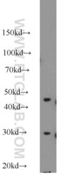
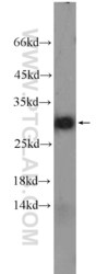
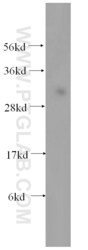
- HEK-293 cells were subjected to SDS PAGE followed by western blot with 15066-1-AP (NDUFS3 antibody) at dilution of 1:500 incubated at room temperature for 1.5 hours.
- Submitted by
- Invitrogen Antibodies (provider)
- Main image

- Experimental details
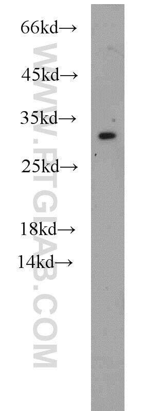
- Human heart tissue were subjected to SDS PAGE followed by western blot with 15066-1-AP (NDUFS3 antibody) at dilution of 1:300 incubated at room temperature for 1.5 hours.
- Submitted by
- Invitrogen Antibodies (provider)
- Main image
- Experimental details
- HEK-293 cells were subjected to SDS PAGE followed by western blot with 15066-1-AP (NDUFS3 antibody) at dilution of 1:500 incubated at room temperature for 1.5 hours.

- Submitted by
- Invitrogen Antibodies (provider)
- Main image
- Experimental details
- HEK-293 cells were subjected to SDS PAGE followed by western blot with 15066-1-AP (NDUFS3 antibody) at dilution of 1:500 incubated at room temperature for 1.5 hours.
- Submitted by
- Invitrogen Antibodies (provider)
- Main image

- Experimental details
- HEK-293 cells were subjected to SDS PAGE followed by western blot with 15066-1-AP (NDUFS3 antibody) at dilution of 1:500 incubated at room temperature for 1.5 hours.
- Submitted by
- Invitrogen Antibodies (provider)
- Main image
- Experimental details
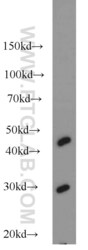
- Mouse heart tissue were subjected to SDS PAGE followed by western blot with 15066-1-AP (NDUFS3 antibody) at dilution of 1:500 incubated at room temperature for 1.5 hours.

- Submitted by
- Invitrogen Antibodies (provider)
- Main image

- Experimental details
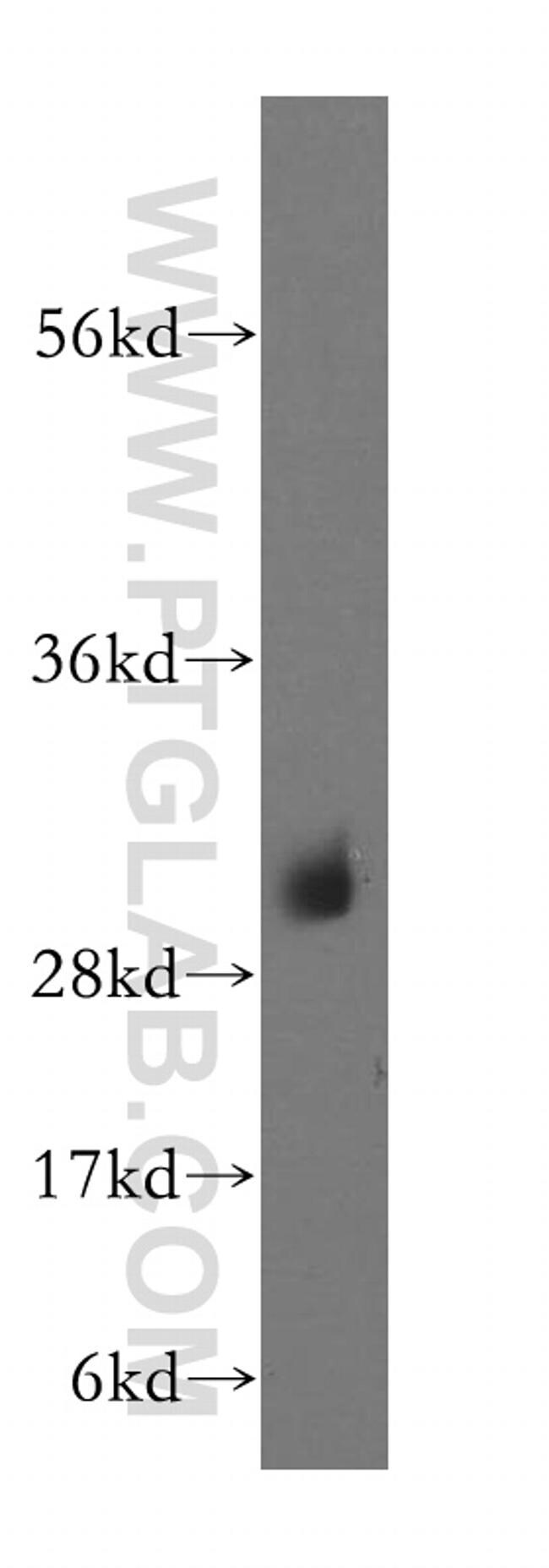
- Mouse skeletal muscle tissue were subjected to SDS PAGE followed by western blot with 15066-1-AP (NDUFS3 antibody) at dilution of 1:400 incubated at room temperature for 1.5 hours.
- Submitted by
- Invitrogen Antibodies (provider)
- Main image
- Experimental details
- HepG2 cells were subjected to SDS PAGE followed by western blot with 15066-1-AP (NDUFS3 antibody) at dilution of 1:300 incubated at room temperature for 1.5 hours.

Supportive validation
- Submitted by
- Invitrogen Antibodies (provider)
- Main image

- Experimental details
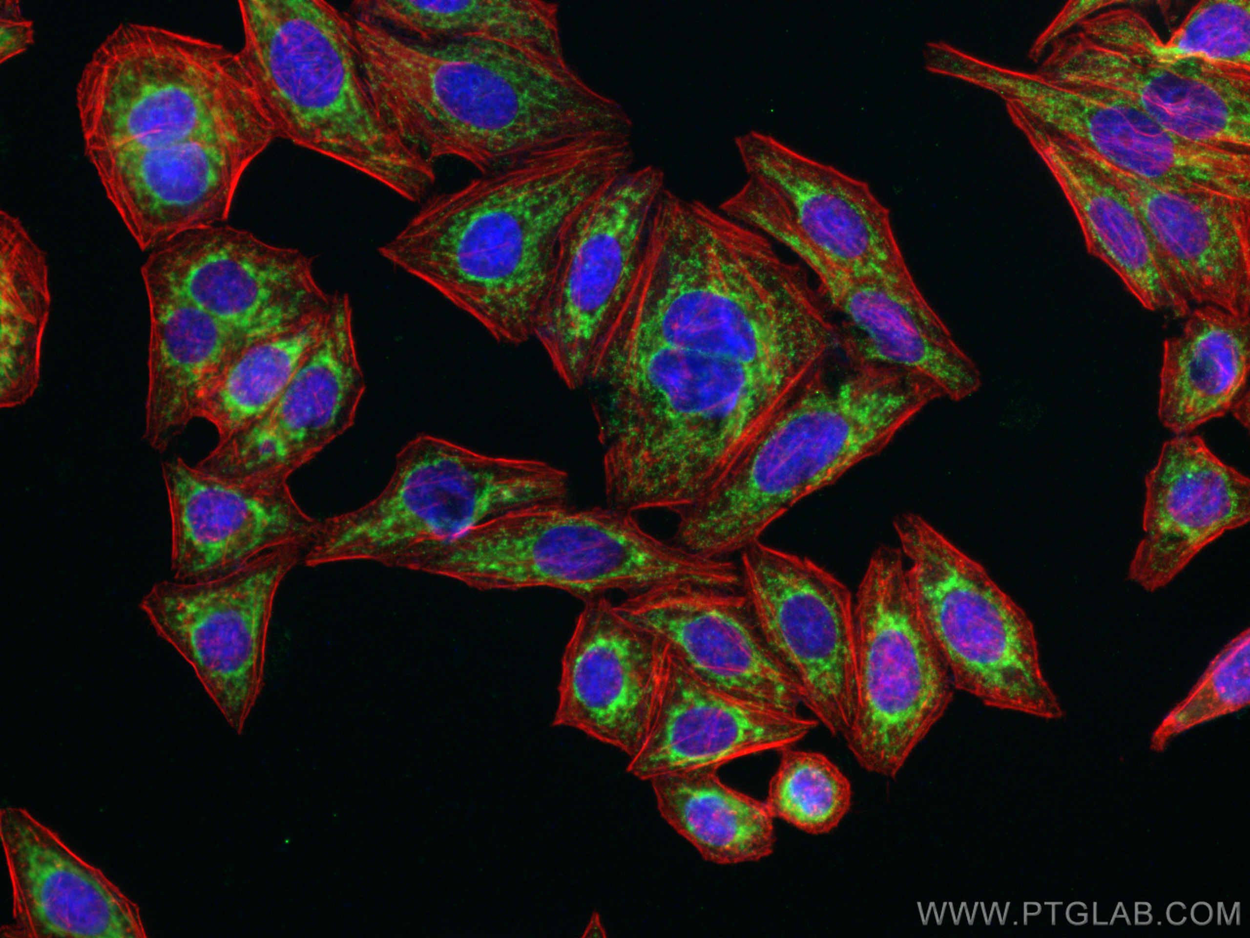
- Immunofluorescent analysis of (-20°C Ethanol) fixed HepG2 cells using NDUFS3 antibody (Product # 15066-1-AP) at dilution of 1:200 and CoraLite®488-Conjugated AffiniPure Goat Anti-Rabbit IgG(H+L).
- Submitted by
- Invitrogen Antibodies (provider)
- Main image
- Experimental details
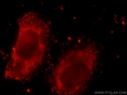
- Immunofluorescent analysis of HepG2 cells using 15066-1-AP (NDUFS3 antibody) at dilution of 1:25 and Rhodamine-Goat anti-Rabbit IGG.

Supportive validation
- Submitted by
- Invitrogen Antibodies (provider)
- Main image

- Experimental details
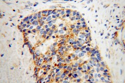
- Immunohistochemistry of paraffin-embedded human liver cancer using 15066-1-AP (NDUFS3 antibody) at dilution of 1:50 (under 10x lens).

- Submitted by
- Invitrogen Antibodies (provider)
- Main image
- Experimental details
- Immunohistochemistry of paraffin-embedded human liver cancer using 15066-1-AP (NDUFS3 antibody) at dilution of 1:50 (under 40x lens).

- Submitted by
- Invitrogen Antibodies (provider)
- Main image
- Experimental details
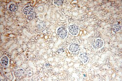
- Immunohistochemistry of paraffin-embedded human kidney using 15066-1-AP (NDUFS3 antibody) at dilution of 1:50 (under 10x lens).

- Submitted by
- Invitrogen Antibodies (provider)
- Main image

- Experimental details
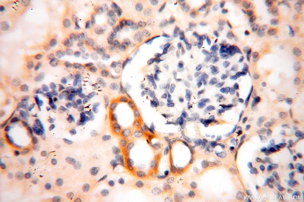
- Immunohistochemistry of paraffin-embedded human kidney using 15066-1-AP (NDUFS3 antibody) at dilution of 1:50 (under 40x lens).
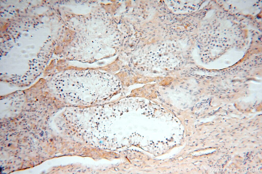
- Submitted by
- Invitrogen Antibodies (provider)
- Main image
- Experimental details
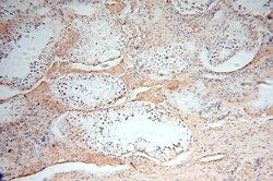
- Immunohistochemistry of paraffin-embedded human testis using 15066-1-AP (NDUFS3 antibody) at dilution of 1:50 (under 10x lens).
- Submitted by
- Invitrogen Antibodies (provider)
- Main image
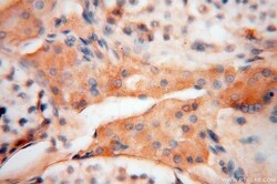
- Experimental details
- Immunohistochemistry of paraffin-embedded human testis using 15066-1-AP (NDUFS3 antibody) at dilution of 1:50 (under 40x lens).

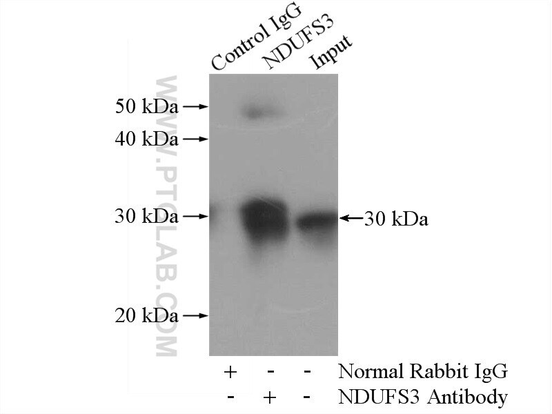
Supportive validation
- Submitted by
- Invitrogen Antibodies (provider)
- Main image

- Experimental details
- IP result of anti-NDUFS3 (IP:15066-1-AP, 4ug; Detection:15066-1-AP 1:500) with mouse heart tissue lysate 4000ug.