Explore
 Validate
 Learn
Learn Immunohistochemistry
Antibody data
- Antibody Data
- Antigen structure
- References [3]
- Comments [0]
- Validations
- Immunohistochemistry [1]
Submit
Validation data
Reference
Comment
Report error
- Product number
- MAB8166 - Provider product page

- Provider
- Abnova Corporation
- Proper citation
- Abnova Corporation Cat#MAB8166, RRID:AB_10678457
- Product name
- LGR4 monoclonal antibody, clone 6G8.B3.G5.C3
- Antibody type
- Monoclonal
- Description
- Mouse monoclonal antibody raised against synthetic peptide of LGR4 .
- Antibody clone number
- 6G8.B3.G5.C3
- Storage
- Store at 4°C. For long term storage store at -20°C.Aliquot to avoid repeated freezing and thawing.
Submitted references Deletion of G protein-coupled receptor 48 leads to ocular anterior segment dysgenesis (ASD) through down-regulation of Pitx2.
Up-regulation of GPR48 induced by down-regulation of p27Kip1 enhances carcinoma cell invasiveness and metastasis.
Molecular characterization of a novel glycoprotein hormone G-protein-coupled receptor.
Weng J, Luo J, Cheng X, Jin C, Zhou X, Qu J, Tu L, Ai D, Li D, Wang J, Martin JF, Amendt BA, Liu M
Proceedings of the National Academy of Sciences of the United States of America 2008 Apr 22;105(16):6081-6
Proceedings of the National Academy of Sciences of the United States of America 2008 Apr 22;105(16):6081-6
Up-regulation of GPR48 induced by down-regulation of p27Kip1 enhances carcinoma cell invasiveness and metastasis.
Gao Y, Kitagawa K, Hiramatsu Y, Kikuchi H, Isobe T, Shimada M, Uchida C, Hattori T, Oda T, Nakayama K, Nakayama KI, Tanaka T, Konno H, Kitagawa M
Cancer research 2006 Dec 15;66(24):11623-31
Cancer research 2006 Dec 15;66(24):11623-31
Molecular characterization of a novel glycoprotein hormone G-protein-coupled receptor.
Loh ED, Broussard SR, Kolakowski LF
Biochemical and biophysical research communications 2001 Apr 6;282(3):757-64
Biochemical and biophysical research communications 2001 Apr 6;282(3):757-64
No comments: Submit comment
Supportive validation
- Submitted by
- Abnova Corporation (provider)
- Main image

- Experimental details
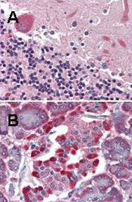
- Immunohistochemical staining of LGR4 monoclonal antibody, clone 6G8.B3.G5.C3 (Cat # MAB8166) was used diluted to 5 ug/mL to detect LGR4 staining at the membrane of cells in various human tissues.A. Brain cerebellum.B. Pancreas islet.Strongly positive staining is noted in subsets of cells within the islets of Langerhans. Moderately positive staiing was observed in Purkinje and Golgi neurons of the cerebellum, adrenal medulla, neuroendocrine cells, hepatocytes, lung macrophages, seminiferous tubules and Leydig cells of the testis. Faintly to moderately positive staining was also observed in cardiac myocytes and renal tubules, granulocytes, and subsets of lymphocytes. Some elastin background staining is noted. Tissue was formalin fixed and paraffin embedded. No pre-treatment of sample was required. The image shows the localization of antibody as the precipitated red signal, with a hematoxylin purple nuclear counterstain.Personal communication, Andrew Elston, Lifespan Biosciences, Seattle, WA.
- Validation comment
- Immunohistochemistry (Formalin/PFA-fixed paraffin-embedded sections)