 Explore
 Validate
 Learn
Learn Western blot
 Immunohistochemistry
Antibody data
- Antibody Data
- Antigen structure
- References [0]
- Comments [0]
- Validations
- Western blot [3]
- Immunocytochemistry [2]
- Immunohistochemistry [1]
Submit
Validation data
Reference
Comment
Report error
- Product number
- HPA041997 - Provider product page

- Provider
- Atlas Antibodies
- Proper citation
- Atlas Antibodies Cat#HPA041997, RRID:AB_10804123
- Product name
- Anti-EIF3G
- Antibody type
- Polyclonal
- Reactivity
- Human
- Host
- Rabbit
- Conjugate
- Unconjugated
- Antigen sequence
ICKGDHWTTRCPYKDTLGPMQKELAEQLGLSTGEK
EKLPGELEPVQATQNKTGKYVPPSLRDGASRRGES
MQPN- Isotype
- IgG
- Vial size
- 100 µl
- Storage
- Store at +4°C for short term storage. Long time storage is recommended at -20°C.
No comments: Submit comment
Enhanced validation
Supportive validation
- Submitted by
- klas2
- Enhanced method
- Genetic validation
- Main image
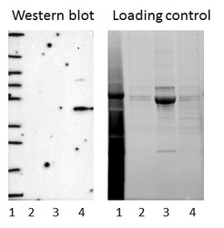
- Experimental details
- Western blot of cell lysate from U-2 OS cells transfected with either siRNA targeting EIF3G or control siRNA. Lane 1: Marker (250, 130, 95, 72, 55, 36, 28, 17, 10) Lane 2: Cell lysate from U-2OS cells transfected with siRNA targeting EIF3G Lane 3: N/A Lane 4: Cell lysate from U-2OS cells transfected with control siRNA Right image, lane 1-4: loading control
- Sample type
- U-2 OS
- Primary Ab dilution
- 1:188
- Conjugate
- Horseradish Peroxidase
- Secondary Ab
- Secondary Ab
- Secondary Ab dilution
- 1:3000
- Knockdown/Genetic Approaches Application
- Western blot

Supportive validation
- Submitted by
- Atlas Antibodies (provider)
- Enhanced method
- Genetic validation
- Main image
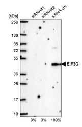
- Experimental details
- Western blot analysis in U2OS cells transfected with control siRNA, target specific siRNA probe #1 and #2, using Anti-EIF3G antibody. Remaining relative intensity is presented.

- Submitted by
- Atlas Antibodies (provider)
- Main image
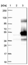
- Experimental details
- Lane 1: Marker [kDa] 250, 130, 95, 72, 55, 36, 28, 17, 10Lane 2: Negative control (vector only transfected HEK293T lysate)Lane 3: Over-expression lysate (Co-expressed with a C-terminal myc-DDK tag (~3.1 kDa) in mammalian HEK293T cells, LY401233)

Enhanced validation
Supportive validation
- Submitted by
- 55af80e3e0991
- Enhanced method
- Genetic validation
- Main image

- Experimental details
- Confocal images of immunofluorescently stained human U-2 OS cells.The protein EIF3G is shown in green. The image to the left show cells transfected with control siRNA and the image to the right show cells where EIF3G has been downregulated with specific siRNA.
- Sample type
- U-2 OS cells
- Primary Ab dilution
- 1:85
- Secondary Ab
- Secondary Ab
- Secondary Ab dilution
- 1:800
- Knockdown/Genetic Approaches Application
- Immunocytochemistry

Supportive validation
- Submitted by
- Atlas Antibodies (provider)
- Main image

- Experimental details
- Immunofluorescent staining of human cell line U-2 OS shows localization to cytosol.
- Sample type
- HUMAN

Supportive validation
- Submitted by
- Atlas Antibodies (provider)
- Main image

- Experimental details
- Immunohistochemical staining of human colon shows strong cytoplasmic positivity in glandular cells.
- Sample type
- HUMAN
