 Explore
 Validate
 Learn
Learn Western blot
Antibody data
- Antibody Data
- Antigen structure
- References [4]
- Comments [0]
- Validations
- Western blot [4]
- Immunocytochemistry [4]
- Immunoprecipitation [1]
- Immunohistochemistry [3]
Submit
Validation data
Reference
Comment
Report error
- Product number
- GTX113241 - Provider product page

- Provider
- GeneTex
- Proper citation
- GeneTex Cat#GTX113241, RRID:AB_1951119
- Product name
- PAX6 antibody
- Antibody type
- Polyclonal
- Reactivity
- Human, Mouse, Rat
- Host
- Rabbit
Submitted references Neural Progenitor-Like Cells Induced from Human Gingiva-Derived Mesenchymal Stem Cells Regulate Myelination of Schwann Cells in Rat Sciatic Nerve Regeneration.
SMN is required for the maintenance of embryonic stem cells and neuronal differentiation in mice.
Role of astroglia in Down's syndrome revealed by patient-derived human-induced pluripotent stem cells.
MicroRNA-328 may influence myopia development by mediating the PAX6 gene.
Zhang Q, Nguyen P, Xu Q, Park W, Lee S, Furuhashi A, Le AD
Stem cells translational medicine 2017 Feb;6(2):458-470
Stem cells translational medicine 2017 Feb;6(2):458-470
SMN is required for the maintenance of embryonic stem cells and neuronal differentiation in mice.
Chang WF, Xu J, Chang CC, Yang SH, Li HY, Hsieh-Li HM, Tsai MH, Wu SC, Cheng WT, Liu JL, Sung LY
Brain structure & function 2015;220(3):1539-53
Brain structure & function 2015;220(3):1539-53
Role of astroglia in Down's syndrome revealed by patient-derived human-induced pluripotent stem cells.
Chen C, Jiang P, Xue H, Peterson SE, Tran HT, McCann AE, Parast MM, Li S, Pleasure DE, Laurent LC, Loring JF, Liu Y, Deng W
Nature communications 2014 Jul 18;5:4430
Nature communications 2014 Jul 18;5:4430
MicroRNA-328 may influence myopia development by mediating the PAX6 gene.
Chen KC, Hsi E, Hu CY, Chou WW, Liang CL, Juo SH
Investigative ophthalmology & visual science 2012 May 31;53(6):2732-9
Investigative ophthalmology & visual science 2012 May 31;53(6):2732-9
No comments: Submit comment
Supportive validation
- Submitted by
- GeneTex (provider)
- Main image
- Experimental details
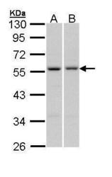
- Sample (30 ?g of whole cell lysate) A: A431 (GTX27909) B: HepG2 (GTX27900) 10% SDS PAGE GTX113241 diluted at 1:1000 The HRP-conjugated anti-rabbit IgG antibody (GTX213110-01) was used to detect the primary antibody.

- Submitted by
- GeneTex (provider)
- Main image

- Experimental details
- Mouse tissue extract (50 ?g) was separated by 10% SDS-PAGE, and the membrane was blotted with PAX6 antibody (GTX113241) diluted at 1:1000. The HRP-conjugated anti-rabbit IgG antibody (GTX213110-01) was used to detect the primary antibody.

- Submitted by
- GeneTex (provider)
- Main image
- Experimental details
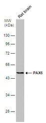
- Rat tissue extract (50 ?g) was separated by 10% SDS-PAGE, and the membrane was blotted with PAX6 antibody (GTX113241) diluted at 1:500. The HRP-conjugated anti-rabbit IgG antibody (GTX213110-01) was used to detect the primary antibody.

- Submitted by
- GeneTex (provider)
- Main image
- Experimental details
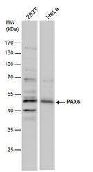
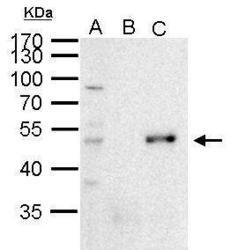
- PAX6 antibody detects PAX6 protein by Western blot analysis. Various whole cell extracts (30 £gg) were separated by 10% SDS-PAGE, and the membrane was blotted with PAX6 antibody (GTX113241) diluted by 1:1000.

Supportive validation
- Submitted by
- GeneTex (provider)
- Main image

- Experimental details
- Immunofluorescence analysis of paraformaldehyde-fixed A549, using PAX6(GTX113241) antibody at 1:200 dilution.

- Submitted by
- GeneTex (provider)
- Main image

- Experimental details
- Immunofluorescence analysis of paraformaldehyde-fixed 293T, using PAX6(GTX113241) antibody at 1:250 dilution.

- Submitted by
- GeneTex (provider)
- Main image
- Experimental details
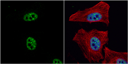
- PAX6 antibody detects PAX6 protein at nucleus by immunofluorescent analysis.Sample: HeLa cells were fixed in 4% paraformaldehyde at RT for 15 min.Green: PAX6 protein stained by PAX6 antibody (GTX113241) diluted at 1:500.Red: alpha Tubulin, a cytoskeleton marker, stained by alpha Tubulin antibody [GT114] (GTX628802) diluted at 1:1000.Blue: Hoechst 33342 staining.

- Submitted by
- GeneTex (provider)
- Main image
- Experimental details
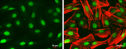
- PAX6 antibody detects PAX6 protein at nucleus by immunofluorescent analysis.Sample: SK-N-SH cells were fixed in 4% paraformaldehyde at RT for 15 min.Green: PAX6 protein stained by PAX6 antibody (GTX113241) diluted at 1:500.Red: Phalloidin, a cytoskeleton marker, diluted at 1:50.Scale bar = 10 £gm.

Supportive validation
- Submitted by
- GeneTex (provider)
- Main image
- Experimental details
- PAX6 antibody immunoprecipitates PAX6 protein in IP experiments. IP Sample: 1000 ?g 293T whole cell lysate/extract A. 50 £gg 293T whole cell lysate/extract B. Control with 2 £gg of preimmune rabbit IgG C. Immunoprecipitation of PAX6 protein by 2 £gg of PAX6 antibody (GTX113241) 10% SDS-PAGE The immunoprecipitated PAX6 protein was detected by PAX6 antibody (GTX113241) diluted at 1:1000. EasyBlot anti-rabbit IgG (GTX221666-01) was used as a secondary reagent.

Supportive validation
- Submitted by
- GeneTex (provider)
- Main image
- Experimental details
- PAX6 antibody detects PAX6 protein at nucleus in embryonic mouse brain by immunohistochemical analysis. Sample: Frozen section of embryonic mouse brain (mE12.5). PAX6 antibody (GTX113241) diluted at 1:250.

- Submitted by
- GeneTex (provider)
- Main image
- Experimental details
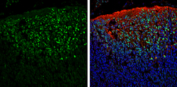
- PAX6 antibody detects PAX6 protein expression by immunohistochemical analysis.Sample: Frozen sectioned E13.5 Rat brain. Green: PAX6 protein stained by PAX6 antibody (GTX113241) diluted at 1:250.Red: beta Tubulin 3/ TUJ1, a mature neuron marker, stained by beta Tubulin 3/ TUJ1 antibody [GT11710] (GTX631836) diluted at 1:500.Blue: Fluoroshield with DAPI (GTX30920).

- Submitted by
- GeneTex (provider)
- Main image
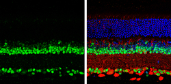
- Experimental details
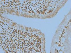
- PAX6 antibody detects PAX6 protein by immunohistochemical analysis.Samples: Paraffin-Embedded mouse retina.Green: PAX6 protein stained by PAX6 antibody (GTX113241) diluted at 1:250.Red: beta Tubulin 3/ Tuj1, stained by beta Tubulin 3/ Tuj1 antibody [GT1338] (GTX631831) diluted at 1:500.Blue: Fluoroshield with DAPI (GTX30920).
