 Explore
 Validate
 Learn
Learn Western blot
Antibody data
- Antibody Data
- Antigen structure
- References [3]
- Comments [0]
- Validations
- Western blot [2]
- Immunocytochemistry [1]
- Flow cytometry [1]
Submit
Validation data
Reference
Comment
Report error
- Product number
- AF8150 - Provider product page

- Provider
- R&D Systems
- Product name
- Human Pax6 Antibody
- Antibody type
- Polyclonal
- Description
- Antigen Affinity-purified. Detects human Pax6 in direct ELISAs. In direct ELISAs, less than 5% cross-reactivity with recombinant human (rh) Pax1, rhPax2, rhPax3, rhPax4, rhPax5, and rhPax7 is observed.
- Reactivity
- Human
- Host
- Sheep
- Conjugate
- Unconjugated
- Antigen sequence
P26367- Isotype
- IgG
- Vial size
- 100 ug
- Concentration
- LYOPH
- Storage
- Use a manual defrost freezer and avoid repeated freeze-thaw cycles. 12 months from date of receipt, -20 to -70 °C as supplied. 1 month, 2 to 8 °C under sterile conditions after reconstitution. 6 months, -20 to -70 °C under sterile conditions after reconstitution.
Submitted references Chronic Exposure to Palmitate Impairs Insulin Signaling in an Intestinal L-cell Line: A Possible Shift from GLP-1 to Glucagon Production.
TFAP2C regulates transcription in human naive pluripotency by opening enhancers.
Generation of nine induced pluripotent stem cell lines as an ethnic diversity panel.
Filippello A, Urbano F, Di Mauro S, Scamporrino A, Di Pino A, Scicali R, Rabuazzo AM, Purrello F, Piro S
International journal of molecular sciences 2018 Nov 28;19(12)
International journal of molecular sciences 2018 Nov 28;19(12)
TFAP2C regulates transcription in human naive pluripotency by opening enhancers.
Pastor WA, Liu W, Chen D, Ho J, Kim R, Hunt TJ, Lukianchikov A, Liu X, Polo JM, Jacobsen SE, Clark AT
Nature cell biology 2018 May;20(5):553-564
Nature cell biology 2018 May;20(5):553-564
Generation of nine induced pluripotent stem cell lines as an ethnic diversity panel.
Gao X, Yourick JJ, Sprando RL
Stem cell research 2018 Aug;31:193-196
Stem cell research 2018 Aug;31:193-196
No comments: Submit comment
Supportive validation
- Submitted by
- R&D Systems (provider)
- Main image
- Experimental details
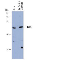
- Detection of Human and Rat Pax6 by Western Blot. Western blot shows lysates of HeLa human cervical epithelial carcinoma cell line and rat cortical stem cells. PVDF membrane was probed with 0.5 µg/mL of Sheep Anti-Human Pax6 Antigen Affinity-purified Polyclonal Antibody (Catalog # AF8150) followed by HRP-conjugated Anti-Sheep IgG Secondary Antibody (Catalog # HAF016). A specific band was detected for Pax6 at approximately 48-50 kDa (as indicated). This experiment was conducted under reducing conditions and using Immunoblot Buffer Group 1.

- Submitted by
- R&D Systems (provider)
- Main image
- Experimental details
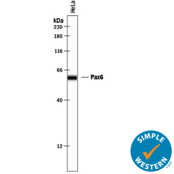
- Detection of Human Pax6 by Simple WesternTM. Simple Western lane view shows lysates of HeLa human cervical epithelial carcinoma cell line, loaded at 0.2 mg/mL. A specific band was detected for Pax6 at approximately 59 kDa (as indicated) using 5 µg/mL of Sheep Anti-Human Pax6 Antigen Affinity-purified Polyclonal Antibody (Catalog # AF8150) followed by 1:50 dilution of HRP-conjugated Anti-Sheep IgG Secondary Antibody (Catalog # HAF016). This experiment was conducted under reducing conditions and using the 12-230 kDa separation system.

Supportive validation
- Submitted by
- R&D Systems (provider)
- Main image

- Experimental details
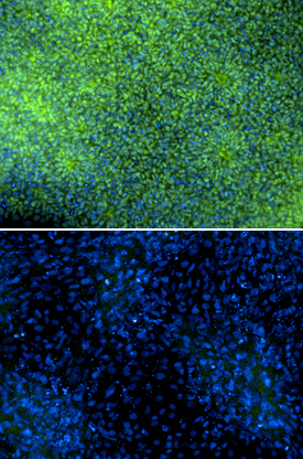
- Pax6 in SA01 Human Embryonic Stem Cells. Immersion fixed SA01 human embryonic stem cells were differentiated for 6 days with Recombinant Human Noggin (Catalog # 6057-NG) and SB431542 (upper panel) or differentiated for 6 days with Recombinant Human BMP-4 (negative control, lower panel; Catalog # 314-BP). Pax6 was detected using Sheep Anti-Human Pax6 Antigen Affinity-purified Polyclonal Antibody (Catalog # AF8150) at 5 µg/mL. Cells were stained using an Alexa Fluor 488-conjugated Anti-Sheep IgG Secondary Antibody (green) and counterstained with DAPI (blue). Specific staining was localized to nuclei. Images courtesy of Dr. Ron McKay, Leiber Institute for Brain Development, Baltimore, Maryland, USA.
Supportive validation
- Submitted by
- R&D Systems (provider)
- Main image
- Experimental details
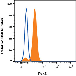
- Detection of Pax6 in HeLa Human Cell Line by Flow Cytometry. HeLa human cervical epithelial carcinoma cell line was stained with Sheep Anti-Human Pax6 Affinity-Purified Polyclonal Antibody (Catalog # AF8150, filled histogram) or Sheep IgG control Antibody (Catalog # 5-001-A, open histogram) followed by anti-Sheep IgG PE-conjugated Secondary Antibody (Catalog # F0126). To facilitate intracellular staining, cells were fixed and permeabilized with FlowX FoxP3 Fixation & Permeabilization Buffer Kit (Catalog # FC012). View our protocol for Staining Intracellular Molecules.
