 Explore
 Validate
 Learn
Learn Western blot
Antibody data
- Antibody Data
- Antigen structure
- References [0]
- Comments [0]
- Validations
- Western blot [3]
- Immunocytochemistry [1]
- Flow cytometry [1]
Submit
Validation data
Reference
Comment
Report error
- Product number
- AF4019 - Provider product page

- Provider
- R&D Systems
- Product name
- Human IRF3 Antibody
- Antibody type
- Polyclonal
- Description
- Antigen Affinity-purified. Detects human IRF3 in direct ELISAs and Western blots. In direct ELISAs, approximately 20% cross-reactivity with recombinant mouse IRF3 is observed.
- Reactivity
- Human
- Host
- Goat
- Conjugate
- Unconjugated
- Antigen sequence
Q14653- Isotype
- IgG
- Vial size
- 100 ug
- Concentration
- LYOPH
- Storage
- Use a manual defrost freezer and avoid repeated freeze-thaw cycles. 12 months from date of receipt, -20 to -70 °C as supplied. 1 month, 2 to 8 °C under sterile conditions after reconstitution. 6 months, -20 to -70 °C under sterile conditions after reconstitution.
No comments: Submit comment
Supportive validation
- Submitted by
- R&D Systems (provider)
- Main image
- Experimental details
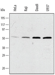
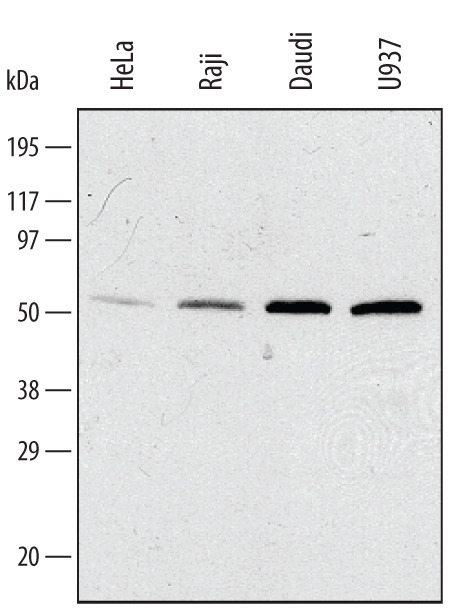
- Detection of Human IRF3 by Western Blot. Western blot shows lysates of HeLa human cervical epithelial carcinoma cell line, U937 human histiocytic lymphoma cell line, Raji human Burkitt's lymphoma cell line, and Daudi human Burkitt's lymphoma cell line. PVDF membrane was probed with 1 µg/mL of Goat Anti-Human IRF3 Antigen Affinity-purified Polyclonal Antibody (Catalog # AF4019) followed by HRP-conjugated Anti-Goat IgG Secondary Antibody (Catalog # HAF019). A specific band was detected for IRF3 at approximately 50 kDa (as indicated). This experiment was conducted under reducing conditions and using Immunoblot Buffer Group 1.
- Submitted by
- R&D Systems (provider)
- Main image

- Experimental details
- Western Blot Shows Human IRF3 Specificity by Using Knockout Cell Line. Western blot shows lysates of HeLa human cervical epithelial carcinoma parental cell line and IRF3 knockout HeLa cell line (KO). PVDF membrane was probed with 1 µg/mL of Goat Anti-Human IRF3 Antigen Affinity-purified Polyclonal Antibody (Catalog # AF4019) followed by HRP-conjugated Anti-Goat IgG Secondary Antibody (Catalog # HAF017). A specific band was detected for IRF3 at approximately 50 kDa (as indicated) in the parental HeLa cell line, but is not detectable in knockout HeLa cell line. GAPDH (Catalog # AF5718) is shown as a loading control. This experiment was conducted under reducing conditions and using Immunoblot Buffer Group 1.

- Submitted by
- R&D Systems (provider)
- Main image
- Experimental details
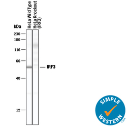
- Detection of Human IRF3 by Simple WesternTM. Simple Western lane view shows lysates of HeLa human cervical epithelial carcinoma parental cell line and IRF3 knockout HeLa cell line (KO), loaded at 0.2 mg/mL. A specific band was detected for IRF3 at approximately 57 kDa (as indicated) in the parental HeLa cell line, but is not detectable in knockout HeLa cell line. Goat Anti-Human IRF3 Antigen Affinity-purified Polyclonal Antibody (Catalog # AF4019) was used at 10 µg/mL followed by 1:50 dilution of HRP-conjugated Anti-Goat IgG Secondary Antibody (Catalog # HAF019). This experiment was conducted under reducing conditions and using the 12-230 kDa separation system.

Supportive validation
- Submitted by
- R&D Systems (provider)
- Main image

- Experimental details
- IRF3 in HeLa Human Cell Line. IRF3 was detected in immersion fixed HeLa human cervical epithelial carcinoma cell line using Goat Anti-Human IRF3 Antigen Affinity-purified Polyclonal Antibody (Catalog # AF4019) at 15 µg/mL for 3 hours at room temperature. Cells were stained using the NorthernLights™ 557-conjugated Anti-Goat IgG Secondary Antibody (red; Catalog # NL001) and counterstained with DAPI (blue). Specific staining was localized to cytoplasm. View our protocol for Fluorescent ICC Staining of Cells on Coverslips.

Supportive validation
- Submitted by
- R&D Systems (provider)
- Main image
- Experimental details
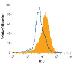
- Detection of IRF3 in Daudi Human Cell Line by Flow Cytometry. Daudi human Burkitt's lymphoma cell line was stained with Goat Anti-Human IRF3 Antigen Affinity-purified Polyclonal Antibody (Catalog # AF4019, filled histogram) or isotype control antibody (Catalog # AB-108-C, open histogram), followed by Phycoerythrin-conjugated Anti-Goat IgG Secondary Antibody (Catalog # F0107). To facilitate intracellular staining, cells were fixed with paraformaldehyde and permeabilized with saponin.
