 Explore
 Validate
 Learn
Learn Western blot
Antibody data
- Antibody Data
- Antigen structure
- References [3]
- Comments [0]
- Validations
- Western blot [3]
- Immunocytochemistry [1]
- Immunoprecipitation [1]
- Immunohistochemistry [1]
Submit
Validation data
Reference
Comment
Report error
- Product number
- GTX103203 - Provider product page

- Provider
- GeneTex
- Proper citation
- GeneTex Cat#GTX103203, RRID:AB_1950517
- Product name
- GRP94 antibody [N1N3]
- Antibody type
- Polyclonal
- Reactivity
- Human, Mouse
- Host
- Rabbit
Submitted references Small molecule proteostasis regulators that reprogram the ER to reduce extracellular protein aggregation.
Serotype O18 avian pathogenic and neonatal meningitis Escherichia coli strains employ similar pathogenic strategies for the onset of meningitis.
Unfolded protein response activation reduces secretion and extracellular aggregation of amyloidogenic immunoglobulin light chain.
Plate L, Cooley CB, Chen JJ, Paxman RJ, Gallagher CM, Madoux F, Genereux JC, Dobbs W, Garza D, Spicer TP, Scampavia L, Brown SJ, Rosen H, Powers ET, Walter P, Hodder P, Wiseman RL, Kelly JW
eLife 2016 Jul 20;5
eLife 2016 Jul 20;5
Serotype O18 avian pathogenic and neonatal meningitis Escherichia coli strains employ similar pathogenic strategies for the onset of meningitis.
Krishnan S, Chang AC, Hodges J, Couraud PO, Romero IA, Weksler B, Nicholson BA, Nolan LK, Prasadarao NV
Virulence 2015;6(8):777-86
Virulence 2015;6(8):777-86
Unfolded protein response activation reduces secretion and extracellular aggregation of amyloidogenic immunoglobulin light chain.
Cooley CB, Ryno LM, Plate L, Morgan GJ, Hulleman JD, Kelly JW, Wiseman RL
Proceedings of the National Academy of Sciences of the United States of America 2014 Sep 9;111(36):13046-51
Proceedings of the National Academy of Sciences of the United States of America 2014 Sep 9;111(36):13046-51
No comments: Submit comment
Supportive validation
- Submitted by
- GeneTex (provider)
- Main image

- Experimental details
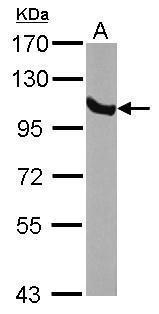
- Sample (30 ug of whole cell lysate) A: NIH-3T3 7.5% SDS PAGE GTX103203 diluted at 1:5000
- Submitted by
- GeneTex (provider)
- Main image
- Experimental details
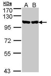
- Sample (30 ug of whole cell lysate)A: H1299B: Hela S37.5% SDS PAGEGTX103203 diluted at 1:1000

- Submitted by
- GeneTex (provider)
- Main image
- Experimental details
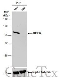
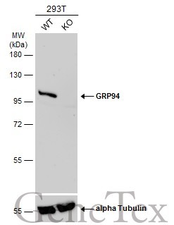
- Wild-type (WT) and GRP94 knockout (KO) 293T cell extracts (30 ?g) were separated by 7.5% SDS-PAGE, and the membrane was blotted with GRP94 antibody [N1N3] (GTX103203) diluted at 1:2000. The HRP-conjugated anti-rabbit IgG antibody (GTX213110-01) was used to detect the primary antibody.
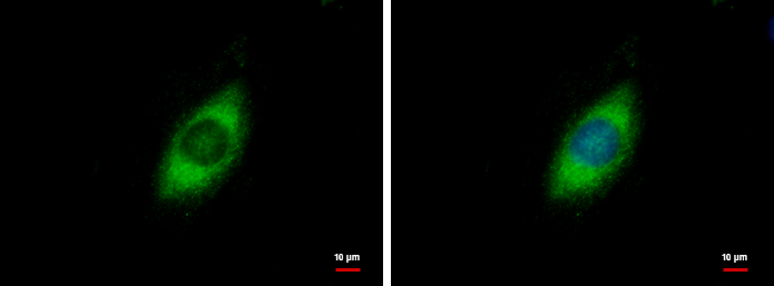
Supportive validation
- Submitted by
- GeneTex (provider)
- Main image

- Experimental details
- GRP94 antibody [N1N3] detects GRP94 protein at cytoplasm by immunofluorescent analysis.Sample: HeLa cells were fixed in 4% paraformaldehyde at RT for 15 min.Green: GRP94 protein stained by GRP94 antibody [N1N3] (GTX103203) diluted at 1:500.Blue: Hoechst 33342 staining.
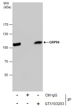
Supportive validation
- Submitted by
- GeneTex (provider)
- Main image
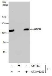
- Experimental details
- Immunoprecipitation of GRP94 protein from HeLa whole cell extracts using 5 £gg of GRP94 antibody [N1N3] (GTX103203).Western blot analysis was performed using GRP94 antibody [N1N3] (GTX103203).EasyBlot anti-Rabbit IgG (GTX221666-01) was used as a secondary reagent.
Supportive validation
- Submitted by
- GeneTex (provider)
- Main image
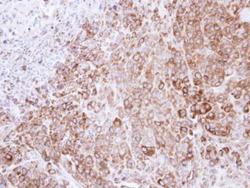
- Experimental details
- Immunohistochemical analysis of paraffin-embedded SCM-1 xenograft , using HSP90B(GTX103203) antibody at 1:500 dilution.
