 Explore
 Validate
 Learn
Learn Western blot
Antibody data
- Antibody Data
- Antigen structure
- References [1]
- Comments [0]
- Validations
- Western blot [2]
- Immunohistochemistry [1]
Submit
Validation data
Reference
Comment
Report error
- Product number
- AF7606 - Provider product page

- Provider
- R&D Systems
- Product name
- Human/Mouse/Rat gp96/HSP90B1 Antibody
- Antibody type
- Polyclonal
- Description
- Antigen Affinity-purified. Detects human, mouse, and rat gp96/HSP90B1 in direct ELISAs and Western blots.
- Reactivity
- Human, Mouse, Rat
- Host
- Sheep
- Conjugate
- Unconjugated
- Antigen sequence
P14625- Isotype
- IgG
- Vial size
- 100 ug
- Concentration
- LYOPH
- Storage
- Use a manual defrost freezer and avoid repeated freeze-thaw cycles. 12 months from date of receipt, -20 to -70 °C as supplied. 1 month, 2 to 8 °C under sterile conditions after reconstitution. 6 months, -20 to -70 °C under sterile conditions after reconstitution.
Submitted references XBP1 signalling is essential for alleviating mutant protein aggregation in ER-stress related skeletal disease.
Piróg KA, Dennis EP, Hartley CL, Jackson RM, Soul J, Schwartz JM, Bateman JF, Boot-Handford RP, Briggs MD
PLoS genetics 2019 Jul;15(7):e1008215
PLoS genetics 2019 Jul;15(7):e1008215
No comments: Submit comment
Supportive validation
- Submitted by
- R&D Systems (provider)
- Main image
- Experimental details
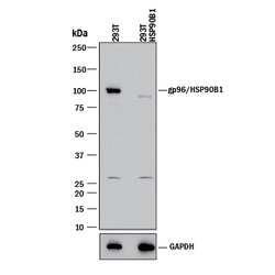
- Western Blot Shows Human gp96/HSP90B1 Specificity by Using Knockout Cell Line. Western blot shows lysates of HEK293T human embryonic kidney parental cell line and gp96/HSP90B1 knockout HEK293T cell line (KO). PVDF membrane was probed with 0.5 µg/mL of Sheep Anti-Human/Mouse/Rat gp96/HSP90B1 Antigen Affinity-purified Polyclonal Antibody (Catalog # AF7606) followed by HRP-conjugated Anti-Sheep IgG Secondary Antibody (Catalog # HAF016). A specific band was detected for gp96/HSP90B1 at approximately 100 kDa (as indicated) in the parental HEK293T cell line, but is not detectable in knockout HEK293T cell line. GAPDH (Catalog # AF5718) is shown as a loading control. This experiment was conducted under reducing conditions and using Immunoblot Buffer Group 1.

- Submitted by
- R&D Systems (provider)
- Main image
- Experimental details
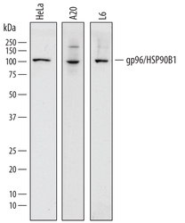
- Detection of Human, Mouse, and Rat gp96/HSP90B1 by Western Blot. Western blot shows lysates of HeLa human cervical epithelial carcinoma cell line, A20 mouse B cell lymphoma cell line, and L6 rat myoblast cell line. PVDF membrane was probed with 0.5 µg/mL of Sheep Anti-Human/Mouse/Rat gp96/HSP90B1 Antigen Affinity-purified Polyclonal Antibody (Catalog # AF7606) followed by HRP-conjugated Anti-Sheep IgG Secondary Antibody (Catalog # HAF016). A specific band was detected for gp96/HSP90B1 at approximately 100 kDa (as indicated). This experiment was conducted under reducing conditions and using Immunoblot Buffer Group 1.

Supportive validation
- Submitted by
- R&D Systems (provider)
- Main image

- Experimental details
- gp96/HSP90B1 in Human Mesothelioma Tissue. gp96/HSP90B1 was detected in immersion fixed paraffin-embedded sections of human mesothelioma tissue using Sheep Anti-Human/Mouse/Rat gp96/HSP90B1 Antigen Affinity-purified Polyclonal Antibody (Catalog # AF7606) at 1 µg/mL overnight at 4 °C. Tissue was stained using the Anti-Sheep HRP-DAB Cell & Tissue Staining Kit (brown; Catalog # CTS019) and counterstained with hematoxylin (blue). Specific staining was localized to cytoplasm and plasma membrane. View our protocol for Chromogenic IHC Staining of Paraffin-embedded Tissue Sections.
