Explore
 Validate
 Learn
Learn Western blot
Antibody data
- Antibody Data
- Antigen structure
- References [1]
- Comments [0]
- Validations
- Western blot [3]
- Immunohistochemistry [5]
- Flow cytometry [2]
Submit
Validation data
Reference
Comment
Report error
- Product number
- TA502698 - Provider product page

- Provider
- OriGene
- Proper citation
- OriGene Cat#TA502698, RRID:AB_11126201
- Product name
- DCK mouse monoclonal antibody, clone OTI16G6 (formerly 16G6)
- Antibody type
- Monoclonal
- Description
- DCK mouse monoclonal antibody, clone OTI16G6 (formerly 16G6)
- Host
- Mouse
- Conjugate
- Unconjugated
- Epitope
- DCK
- Isotype
- IgG
- Antibody clone number
- OTI16G6
- Vial size
- 100 µl
- Concentration
- 1.00mg/ml
Submitted references Prognostic effect of hENT1, dCK and HuR expression by morphological type in periampullary adenocarcinoma, including pancreatic cancer.
Elebro J, Ben Dror L, Heby M, Nodin B, Jirström K, Eberhard J
Acta oncologica (Stockholm, Sweden) 2016;55(3):286-96
Acta oncologica (Stockholm, Sweden) 2016;55(3):286-96
No comments: Submit comment
Supportive validation
- Submitted by
- OriGene (provider)
- Main image

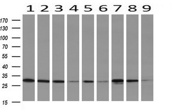
- Experimental details
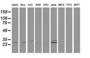
- Western blot analysis of extracts (35ug) from 9 different cell lines by using anti-DCK monoclonal antibody (HepG2: human; HeLa: human; SVT2: mouse; A549: human; COS7: monkey; Jurkat: human; MDCK: canine; PC12: rat; MCF7: human).
- Validation comment
- WB
- Submitted by
- OriGene (provider)
- Main image
- Experimental details
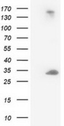
- HEK293T cells were transfected with the pCMV6-ENTRY control (Left lane) or pCMV6-ENTRY DCK (RC210767, Right lane) cDNA for 48 hrs and lysed. Equivalent amounts of cell lysates (5 ug per lane) were separated by SDS-PAGE and immunoblotted with anti-DCK.
- Validation comment
- WB

- Submitted by
- OriGene (provider)
- Main image
- Experimental details
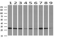
- Western blot analysis of extracts (10ug) from 9 Human tissue by using anti-DCK monoclonal antibody at 1:200 (1: Testis; 2: Omentum; 3: Uterus; 4: Breast; 5: Brain; 6: Liver; 7: Ovary; 8: Thyroid gland; 9: colon).
- Validation comment
- WB
Supportive validation
- Submitted by
- OriGene (provider)
- Main image
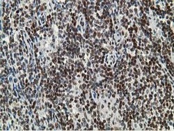
- Experimental details
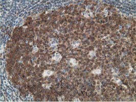
- Immunohistochemical staining of paraffin-embedded Human lymphoma tissue using anti-DCK mouse monoclonal antibody. (Heat-induced epitope retrieval by 10mM citric buffer, pH6.0, 100C for 10min, TA502698)
- Validation comment
- IHC
- Submitted by
- OriGene (provider)
- Main image

- Experimental details
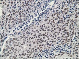
- Immunohistochemical staining of paraffin-embedded Carcinoma of Human lung tissue using anti-DCK mouse monoclonal antibody. (Heat-induced epitope retrieval by 10mM citric buffer, pH6.0, 100C for 10min, TA502698)
- Validation comment
- IHC
- Submitted by
- OriGene (provider)
- Main image

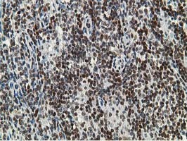
- Experimental details
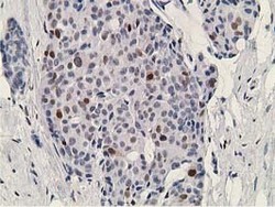
- Immunohistochemical staining of paraffin-embedded Human lymph node tissue within the normal limits using anti-DCK mouse monoclonal antibody. (Heat-induced epitope retrieval by 10mM citric buffer, pH6.0, 100C for 10min, TA502698)
- Validation comment
- IHC
- Submitted by
- OriGene (provider)
- Main image

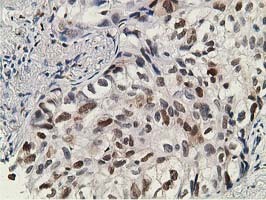
- Experimental details
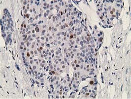
- Immunohistochemical staining of paraffin-embedded Carcinoma of Human bladder tissue using anti-DCK mouse monoclonal antibody. (Heat-induced epitope retrieval by 10mM citric buffer, pH6.0, 100C for 10min, TA502698)
- Validation comment
- IHC
- Submitted by
- OriGene (provider)
- Main image
- Experimental details
- Immunohistochemical staining of paraffin-embedded Adenocarcinoma of Human breast tissue using anti-DCK mouse monoclonal antibody. (Heat-induced epitope retrieval by 10mM citric buffer, pH6.0, 100C for 10min, TA502698)
- Validation comment
- IHC
Supportive validation
- Submitted by
- OriGene (provider)
- Main image
- Experimental details
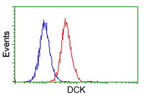
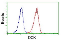
- Flow cytometric Analysis of Jurkat cells, using anti-DCK antibody(TA502698),(Red), compared to a nonspecific negative control antibody,(Blue).
- Validation comment
- FC
- Submitted by
- OriGene (provider)
- Main image
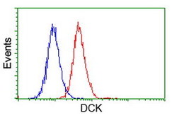
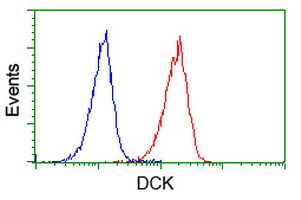
- Experimental details
- Flow cytometric Analysis of Hela cells, using anti-DCK antibody(TA502698),(Red), compared to a nonspecific negative control antibody,(Blue).
- Validation comment
- FC