 Explore
 Validate
 Learn
Learn Western blot
Antibody data
- Antibody Data
- Antigen structure
- References [1]
- Comments [0]
- Validations
- Western blot [1]
- Immunocytochemistry [3]
- Immunohistochemistry [2]
- Flow cytometry [1]
- Other assay [3]
Submit
Validation data
Reference
Comment
Report error
- Product number
- MA5-32331 - Provider product page

- Provider
- Invitrogen Antibodies
- Product name
- Cyclin H Recombinant Rabbit Monoclonal Antibody (SN20-48)
- Antibody type
- Monoclonal
- Antigen
- Recombinant full-length protein
- Reactivity
- Human, Mouse
- Host
- Rabbit
- Isotype
- IgG
- Antibody clone number
- SN20-48
- Vial size
- 100 µL
- Concentration
- 1 mg/mL
- Storage
- Store at 4°C short term. For long term storage, store at -20°C, avoiding freeze/thaw cycles.
Submitted references Cyclin H predicts the poor prognosis and promotes the proliferation of ovarian cancer.
Peng C, Yang Y, Ji L, Yang P, Yang X, Zhang Y
Cancer cell international 2020;20:316
Cancer cell international 2020;20:316
No comments: Submit comment
Supportive validation
- Submitted by
- Invitrogen Antibodies (provider)
- Main image

- Experimental details
- Western blot analysis of Cyclin H in K562 cell lysate using a Cyclin H Monoclonal antibody (Product # MA5-32331) at a dilution of 1:1,000.

Supportive validation
- Submitted by
- Invitrogen Antibodies (provider)
- Main image

- Experimental details
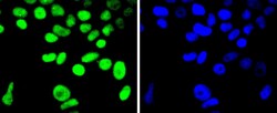
- Immunocytochemical analysis of Cyclin H in Hela cells using a Cyclin H Monoclonal antibody (Product # MA5-32331) as seen in green. The nuclear counter stain is DAPI (blue). Cells were fixed in paraformaldehyde, permeabilised with 0.25% Triton X100/PBS.

- Submitted by
- Invitrogen Antibodies (provider)
- Main image
- Experimental details
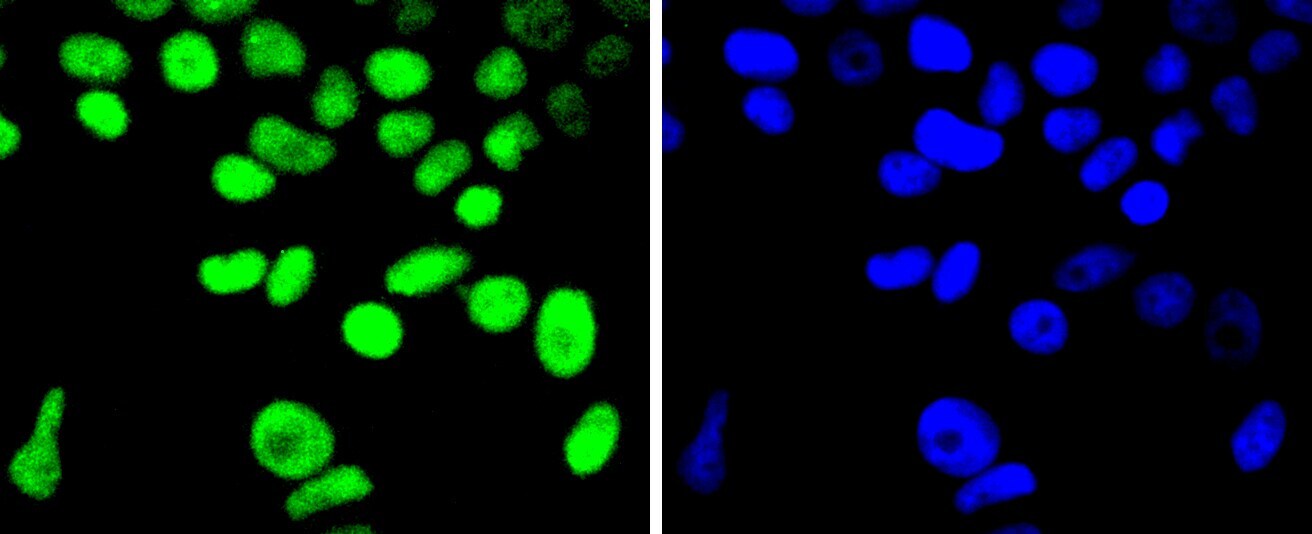
- Immunocytochemical analysis of Cyclin H in MCF-7 cells using a Cyclin H Monoclonal antibody (Product # MA5-32331) as seen in green. The nuclear counter stain is DAPI (blue). Cells were fixed in paraformaldehyde, permeabilised with 0.25% Triton X100/PBS.
- Submitted by
- Invitrogen Antibodies (provider)
- Main image

- Experimental details
- Immunocytochemical analysis of Cyclin H in PC-3M cells using a Cyclin H Monoclonal antibody (Product # MA5-32331) as seen in green. The nuclear counter stain is DAPI (blue). Cells were fixed in paraformaldehyde, permeabilised with 0.25% Triton X100/PBS.

Supportive validation
- Submitted by
- Invitrogen Antibodies (provider)
- Main image
- Experimental details
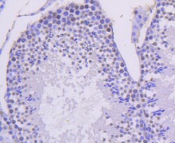
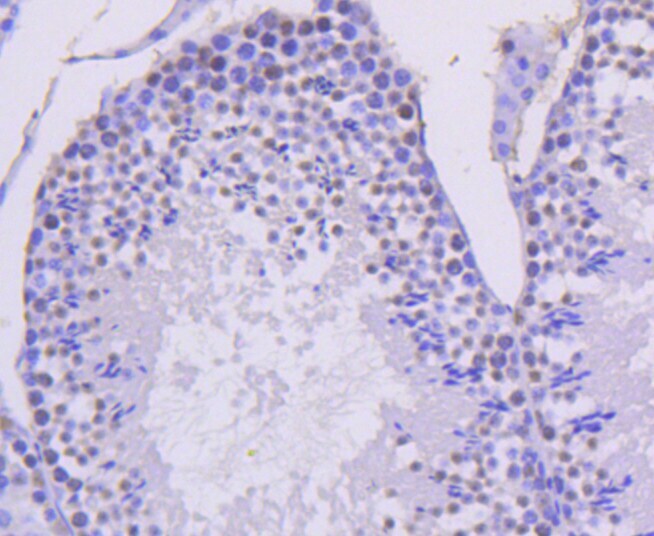
- Immunohistochemical analysis of Cyclin H of paraffin-embedded Mouse testis tissue using a Cyclin-H Monoclonal antibody (Product #MA5-32331). Counter stained with hematoxylin.
- Submitted by
- Invitrogen Antibodies (provider)
- Main image
- Experimental details
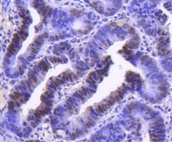
- Immunohistochemical analysis of Cyclin H of paraffin-embedded Human colon cancer tissue using a Cyclin-H Monoclonal antibody (Product #MA5-32331). Counter stained with hematoxylin.

Supportive validation
- Submitted by
- Invitrogen Antibodies (provider)
- Main image

- Experimental details
- Flow Cytometric analysis of Cyclin H in Hela cells using a Cyclin H Monoclonal Antibody (Product # MA5-32331) at a dilution of 1:50, as seen in red compared with an unlabelled control (cells without incubation with primary antibody; black). Alexa Fluor 488-conjugated goat anti rabbit IgG was used as the secondary antibody.

Supportive validation
- Submitted by
- Invitrogen Antibodies (provider)
- Main image

- Experimental details
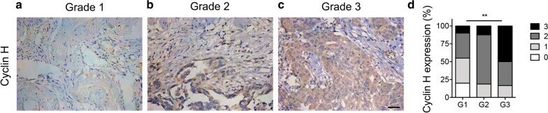
- Fig. 1 Immunohistochemistry staining of Cyclin H in ovarian cancer tissues. The expression of Cyclin H in ovarian cancer of grade 1 ( a ), grade 2 ( b ), and grade 3 ( c ) was measured by immunohistochemistry staining, and representative images are displayed. Scale bar, 20 um. d Quantification of Cyclin H expression in ovarian cancer (P < 0.001)
- Submitted by
- Invitrogen Antibodies (provider)
- Main image
- Experimental details
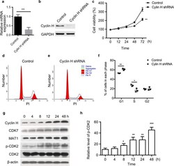
- Fig. 4 Cyclin H regulates the proliferation and cell cycle of ovarian cancer cells. a mRNA level of cyclin H in vector and cyclin H shRNA transfected ovarian cancer HO8910 cells. b The protein expression of cyclin H was detected by Western blot. c Proliferation difference between control and cyclin H silencing HO8910 cells. PI staining was performed to measure the percentage of cells in each cell cycle phase using Modfit software ( d and e ). f Quantification of the percentage cells in each cell cycle phase after transfection with cyclin H shRNA. g Expression of cyclin H, CDK7, MAT1, p-CDK2, and CDK2 in HO8910 cells after serum deprivation and refeeding. Serum-starved HO8910 cells were cultured in serum-containing medium for 4, 8, 12, 24, and 48 h, and cell lysates were analyzed by western blot. h Relative level of p-CDK2 was normalized to the total level of CDK2 at each time point. * P < 0.05, **P < 0.01, ***P < 0.001

- Submitted by
- Invitrogen Antibodies (provider)
- Main image

- Experimental details
- Additional file 1: Figure S1. Expression of cyclin H in different ovarian cancer cell lines. The protein levels of cyclin H in HO8910, SK-OV-3, and NIH: OVCAR-3 cells were detected by western blot. Cyclin H was highly expressed in HO8910 cells.
