Explore
 Validate
 Learn
Learn Western blot
Antibody data
- Antibody Data
- Antigen structure
- References [1]
- Comments [0]
- Validations
- Western blot [3]
- Immunocytochemistry [1]
- Immunohistochemistry [2]
- Other assay [2]
Submit
Validation data
Reference
Comment
Report error
- Product number
- PA5-34687 - Provider product page

- Provider
- Invitrogen Antibodies
- Product name
- BIN1 Polyclonal Antibody
- Antibody type
- Polyclonal
- Antigen
- Recombinant protein fragment
- Reactivity
- Human, Mouse
- Host
- Rabbit
- Isotype
- IgG
- Vial size
- 100 µL
- Concentration
- 2.35 mg/mL
- Storage
- Store at 4°C short term. For long term storage, store at -20°C, avoiding freeze/thaw cycles.
Submitted references Upregulation of RIN3 induces endosomal dysfunction in Alzheimer's disease.
Shen R, Zhao X, He L, Ding Y, Xu W, Lin S, Fang S, Yang W, Sung K, Spencer B, Rissman RA, Lei M, Ding J, Wu C
Translational neurodegeneration 2020 Jun 18;9(1):26
Translational neurodegeneration 2020 Jun 18;9(1):26
No comments: Submit comment
Supportive validation
- Submitted by
- Invitrogen Antibodies (provider)
- Main image
- Experimental details
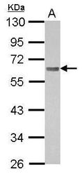
- Western blot analysis of BIN1 using 30 µg of A431 lysate. Samples were loaded onto a 10% SDS-PAGE gel and probed with a BIN1 polyclonal antibody (Product # PA5-34687) at a dilution of 1:1000.

- Submitted by
- Invitrogen Antibodies (provider)
- Main image
- Experimental details
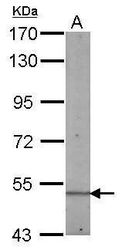
- Western blot analysis of BIN1 using 50 µg of mouse kidney lysate. Samples were loaded onto a 7.5% SDS-PAGE gel and probed with a BIN1 polyclonal antibody (Product # PA5-34687) at a dilution of 1:500.
- Submitted by
- Invitrogen Antibodies (provider)
- Main image
- Experimental details
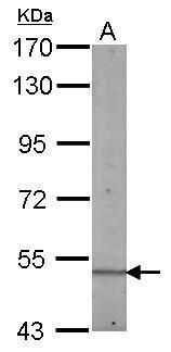
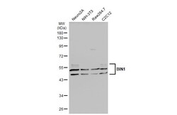
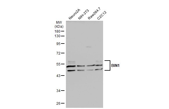
- Western Blot analysis of BIN1 was performed by separating 30 µg of various whole cell extracts by 10% SDS-PAGE. Proteins were transferred to a membrane and probed with a BIN1 Polyclonal Antibody (Product # PA5-34687) at a dilution of 1:2000 and a HRP-conjugated anti-rabbit IgG secondary antibody.
Supportive validation
- Submitted by
- Invitrogen Antibodies (provider)
- Main image
- Experimental details
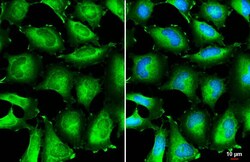
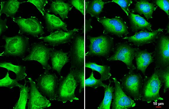
- BIN1 Polyclonal Antibody detects BIN1 protein at cytoplasm by immunofluorescent analysis. Sample: HeLa cells were fixed in 4% paraformaldehyde at RT for 15 min. Green: BIN1 stained by BIN1 Polyclonal Antibody (Product # PA5-34687) diluted at 1:500. Blue: Fluoroshield with DAPI . Scale bar= 10 µm.
Supportive validation
- Submitted by
- Invitrogen Antibodies (provider)
- Main image
- Experimental details
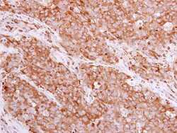
- Immunohistochemistry (Paraffin) analysis of BIN1 was performed in paraffin-embedded human papillary adenocarcinoma tissue using BIN1 Polyclonal Antibody (Product # PA5-34687) at a dilution of 1:500.

- Submitted by
- Invitrogen Antibodies (provider)
- Main image
- Experimental details
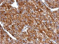
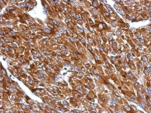
- Immunohistochemical analysis of paraffin-embedded U87 xenograft, using BIN1 (Product # PA5-34687) antibody at 1:500 dilution. Antigen Retrieval: EDTA based buffer, pH 8.0, 15 min.
Supportive validation
- Submitted by
- Invitrogen Antibodies (provider)
- Main image
- Experimental details
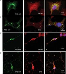
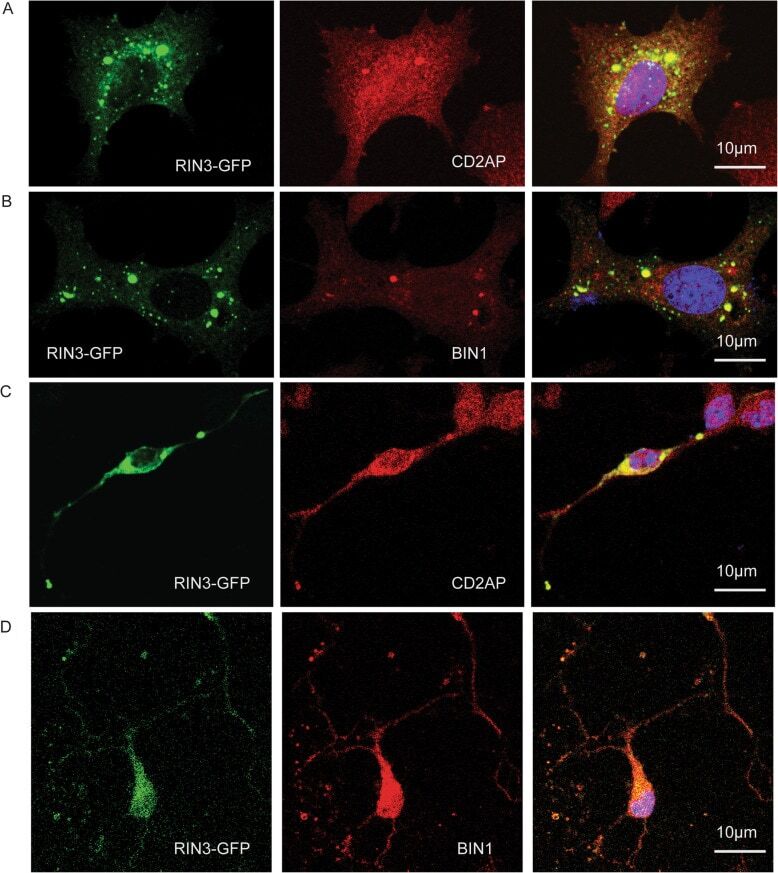
- Fig. 4 RIN3 colocalizes with CD2AP and BIN1. PC12M cells ( a and b ) and primary cortical neurons ( c and d ) were transfected with RIN3-GFP, followed by immunostaining for CD2AP ( a , c ) or BIN1 ( b , d ) or CD2AP using specific antibodies. Yellow color denotes colocalization. Representative images are shown
- Submitted by
- Invitrogen Antibodies (provider)
- Main image

- Experimental details
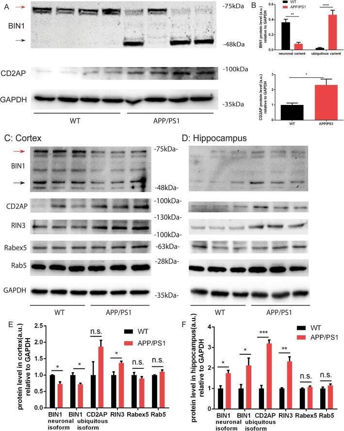
- Fig. 7 BIN1 and CD2AP expression level in APP/PS1 mice. 3-month-old APP/PS1 mice and age-matched WT mice were dissected, lysates were immunoblotted for BIN1 and CD2AP ( a ). The protein levels were normalized against GAPDH as a loading control ( b ). In Cortex ( c ) and hippocampus ( d ) were also extracted from10-month-old APP/PS1 and age-matched WT mice. Protein lysates were used to probe for BIN1, CD2AP, RIN3, Rabex5 and Rab5 using indicated antibodies by SDS-PAGE/immunoblotting ( c and d ), The intensity of bands was quantitated using BioRad-ImageLab. The respective protein levels were normalized against GAPDH as a loading control ( e , f ). p < 0.05 (*), p < 0.01(**), p < 0.0001 (****), standard t-test