Explore
 Validate
 Learn
LearnMA5-11202
antibody from Invitrogen Antibodies
Targeting: MUC1
ADMCKD, ADMCKD1, CD227, MCD, MCKD, MCKD1, PEM, PUM
 Western blot
Antibody data
- Antibody Data
- Antigen structure
- References [59]
- Comments [0]
- Validations
- Western blot [3]
- Immunocytochemistry [5]
- Immunohistochemistry [2]
- Flow cytometry [3]
- Other assay [9]
Submit
Validation data
Reference
Comment
Report error
- Product number
- MA5-11202 - Provider product page

- Provider
- Invitrogen Antibodies
- Product name
- MUC1 Monoclonal Antibody (MH1 (CT2))
- Antibody type
- Monoclonal
- Antigen
- Synthetic peptide
- Description
- MA5-11202 detects smaller subunits (14-28kDa) of MUC-1. Muc-1 is overexpressed in Breast cancer cells. T47D cell lysate could be used as a positive control.
- Reactivity
- Human, Mouse
- Isotype
- IgG
- Antibody clone number
- MH1 (CT2)
- Vial size
- 500 µL
- Concentration
- 0.1 mg/mL
- Storage
- 4° C
Submitted references MUC1-mediated Macrophage Activation Promotes Colitis-associated Colorectal Cancer via Activating the Interleukin-6/ Signal Transducer and Activator of Transcription 3 Axis.
Membrane-Tethered Mucin 1 Is Stimulated by Interferon and Virus Infection in Multiple Cell Types and Inhibits Influenza A Virus Infection in Human Airway Epithelium.
Asynchrony of Apical Polarization, Luminogenesis, and Functional Differentiation in the Developing Thyroid Gland.
Apical Restriction of the Planar Cell Polarity Component VANGL in Pancreatic Ducts Is Required to Maintain Epithelial Integrity.
Jag1 Modulates an Oscillatory Dll1-Notch-Hes1 Signaling Module to Coordinate Growth and Fate of Pancreatic Progenitors.
Nrf2 activation protects against lithium-induced nephrogenic diabetes insipidus.
MUC1 downregulation promotes TNF-α-induced necroptosis in human bronchial epithelial cells via regulation of the RIPK1/RIPK3 pathway.
Essential Role of Protein Arginine Methyltransferase 1 in Pancreas Development by Regulating Protein Stability of Neurogenin 3.
Regulatory effect of dexamethasone on tracheal calcium processing proteins and mucosal secretion.
A Sox2-Sox9 signalling axis maintains human breast luminal progenitor and breast cancer stem cells.
Generation and phenotypic characterization of Pde1a mutant mice.
Targeting MUC1-C inhibits the AKT-S6K1-elF4A pathway regulating TIGAR translation in colorectal cancer.
Recurrent intracranial neurenteric cyst with malignant transformation: A case report and literature review.
MUC1 stimulates EGFR expression and function in endometrial cancer.
Spontaneous Pancreatitis Caused by Tissue-Specific Gene Ablation of Hhex in Mice.
Micro-RNAs miR-29a and miR-330-5p function as tumor suppressors by targeting the MUC1 mucin in pancreatic cancer cells.
Mucin1 shifts Smad3 signaling from the tumor-suppressive pSmad3C/p21(WAF1) pathway to the oncogenic pSmad3L/c-Myc pathway by activating JNK in human hepatocellular carcinoma cells.
Transcription factors SOX4 and SOX9 cooperatively control development of bile ducts.
The chromatin regulator Brg1 suppresses formation of intraductal papillary mucinous neoplasm and pancreatic ductal adenocarcinoma.
MUC1-C oncoprotein activates the ZEB1/miR-200c regulatory loop and epithelial-mesenchymal transition.
Efficient generation of lung and airway epithelial cells from human pluripotent stem cells.
MUC1-C oncoprotein promotes FLT3 receptor activation in acute myeloid leukemia cells.
MUC1 in macrophage: contributions to cigarette smoke-induced lung cancer.
MUC1-C nuclear localization drives invasiveness of renal cancer cells through a sheddase/gamma secretase dependent pathway.
Elimination of von Hippel-Lindau function perturbs pancreas endocrine homeostasis in mice.
MUC1 enhances hypoxia-driven angiogenesis through the regulation of multiple proangiogenic factors.
MUC1-C oncoprotein activates ERK→C/EBPβ signaling and induction of aldehyde dehydrogenase 1A1 in breast cancer cells.
Canonical Notch2 signaling determines biliary cell fates of embryonic hepatoblasts and adult hepatocytes independent of Hes1.
Expression of human full-length MUC1 inhibits the proliferation and migration of a B16 mouse melanoma cell line.
Oncogenic MUC1-C promotes tamoxifen resistance in human breast cancer.
Disrupting BCR-ABL in combination with secondary leukemia-specific pathways in CML cells leads to enhanced apoptosis and decreased proliferation.
Lung cancer with loss of BRG1/BRM, shows epithelial mesenchymal transition phenotype and distinct histologic and genetic features.
Expression of underglycosylated MUC1 antigen in cancerous and adjacent normal breast tissues.
Targeting the eIF4A RNA helicase blocks translation of the MUC1-C oncoprotein.
The MUC1 extracellular domain subunit is found in nuclear speckles and associates with spliceosomes.
Cooperative interaction between the MUC1-C oncoprotein and the Rab31 GTPase in estrogen receptor-positive breast cancer cells.
A novel anti-MUC1 antibody against the MUC1 cytoplasmic tail domain: use in sensitive identification of poorly differentiated cells in adenocarcinoma of the stomach.
GATA4 and GATA6 control mouse pancreas organogenesis.
MUC1-C oncoprotein induces TCF7L2 transcription factor activation and promotes cyclin D1 expression in human breast cancer cells.
Role of the ductal transcription factors HNF6 and Sox9 in pancreatic acinar-to-ductal metaplasia.
Intrinsic mitochondrial membrane potential and associated tumor phenotype are independent of MUC1 over-expression.
MUC1-C oncoprotein regulates glycolysis and pyruvate kinase M2 activity in cancer cells.
MUC1-C oncoprotein suppresses reactive oxygen species-induced terminal differentiation of acute myelogenous leukemia cells.
A mouse model for monitoring islet cell genesis and developing therapies for diabetes.
MUC1-C oncoprotein promotes STAT3 activation in an autoinductive regulatory loop.
Nm23-h1 indirectly promotes the survival of acute myeloid leukemia blast cells by binding to more mature components of the leukemic clone.
Cooperativity of the MUC1 oncoprotein and STAT1 pathway in poor prognosis human breast cancer.
Exocrine-to-endocrine differentiation is detectable only prior to birth in the uninjured mouse pancreas.
MUC1-C oncoprotein functions as a direct activator of the nuclear factor-kappaB p65 transcription factor.
MUC1, a new hypoxia inducible factor target gene, is an actor in clear renal cell carcinoma tumor progression.
Hypoxia enhances MUC1 expression in a lung adenocarcinoma cell line.
A minimal fragment of MUC1 mediates growth of cancer cells.
MUC1 oncoprotein activates the IkappaB kinase beta complex and constitutive NF-kappaB signalling.
Diagnostic value of mucins (MUC1, MUC2 and MUC5AC) expression profile in endoscopic ultrasound-guided fine-needle aspiration specimens of the pancreas.
Multiple tumor types may originate from bone marrow-derived cells.
Localization of putative stem cells and four cell populations with different differentiation degree in mouse mammary anlagen.
Dendritic cells induce MUC1 expression and polarization on human T cells by an IL-7-dependent mechanism.
Dendritic cells induce MUC1 expression and polarization on human T cells by an IL-7-dependent mechanism.
MUC1 oncoprotein activates the FOXO3a transcription factor in a survival response to oxidative stress.
Sheng YH, Davies JM, Wang R, Wong KY, Giri R, Yang Y, Begun J, Florin TH, Hasnain SZ, McGuckin MA
Cellular and molecular gastroenterology and hepatology 2022;14(4):789-811
Cellular and molecular gastroenterology and hepatology 2022;14(4):789-811
Membrane-Tethered Mucin 1 Is Stimulated by Interferon and Virus Infection in Multiple Cell Types and Inhibits Influenza A Virus Infection in Human Airway Epithelium.
Iverson E, Griswold K, Song D, Gagliardi TB, Hamidzadeh K, Kesimer M, Sinha S, Perry M, Duncan GA, Scull MA
mBio 2022 Aug 30;13(4):e0105522
mBio 2022 Aug 30;13(4):e0105522
Asynchrony of Apical Polarization, Luminogenesis, and Functional Differentiation in the Developing Thyroid Gland.
Johansson E, Liang S, Moccia C, Carlsson T, Andersson D, Fagman H, Nilsson M
Frontiers in endocrinology 2021;12:760541
Frontiers in endocrinology 2021;12:760541
Apical Restriction of the Planar Cell Polarity Component VANGL in Pancreatic Ducts Is Required to Maintain Epithelial Integrity.
Flasse L, Yennek S, Cortijo C, Barandiaran IS, Kraus MR, Grapin-Botton A
Cell reports 2020 May 26;31(8):107677
Cell reports 2020 May 26;31(8):107677
Jag1 Modulates an Oscillatory Dll1-Notch-Hes1 Signaling Module to Coordinate Growth and Fate of Pancreatic Progenitors.
Seymour PA, Collin CA, Egeskov-Madsen AR, Jørgensen MC, Shimojo H, Imayoshi I, de Lichtenberg KH, Kopan R, Kageyama R, Serup P
Developmental cell 2020 Mar 23;52(6):731-747.e8
Developmental cell 2020 Mar 23;52(6):731-747.e8
Nrf2 activation protects against lithium-induced nephrogenic diabetes insipidus.
Jobbagy S, Vitturi DA, Salvatore SR, Pires MF, Rowart P, Emlet DR, Ross M, Hahn S, St Croix C, Wendell SG, Subramanya AR, Straub AC, Tan RJ, Schopfer FJ
JCI insight 2020 Jan 16;5(1)
JCI insight 2020 Jan 16;5(1)
MUC1 downregulation promotes TNF-α-induced necroptosis in human bronchial epithelial cells via regulation of the RIPK1/RIPK3 pathway.
Zhang H, Ji J, Liu Q, Xu S
Journal of cellular physiology 2019 Sep;234(9):15080-15088
Journal of cellular physiology 2019 Sep;234(9):15080-15088
Essential Role of Protein Arginine Methyltransferase 1 in Pancreas Development by Regulating Protein Stability of Neurogenin 3.
Lee K, Kim H, Lee J, Oh CM, Song H, Kim H, Koo SH, Lee J, Lim A, Kim H
Diabetes & metabolism journal 2019 Oct;43(5):649-658
Diabetes & metabolism journal 2019 Oct;43(5):649-658
Regulatory effect of dexamethasone on tracheal calcium processing proteins and mucosal secretion.
Lee B, Ahn C, Jeon BH, Jung EM, Yoo YM, Jeung EB
Journal of physiology and pharmacology : an official journal of the Polish Physiological Society 2019 Feb;70(1)
Journal of physiology and pharmacology : an official journal of the Polish Physiological Society 2019 Feb;70(1)
A Sox2-Sox9 signalling axis maintains human breast luminal progenitor and breast cancer stem cells.
Domenici G, Aurrekoetxea-Rodríguez I, Simões BM, Rábano M, Lee SY, Millán JS, Comaills V, Oliemuller E, López-Ruiz JA, Zabalza I, Howard BA, Kypta RM, Vivanco MD
Oncogene 2019 Apr;38(17):3151-3169
Oncogene 2019 Apr;38(17):3151-3169
Generation and phenotypic characterization of Pde1a mutant mice.
Wang X, Yamada S, LaRiviere WB, Ye H, Bakeberg JL, Irazabal MV, Chebib FT, van Deursen J, Harris PC, Sussman CR, Behfar A, Ward CJ, Torres VE
PloS one 2017;12(7):e0181087
PloS one 2017;12(7):e0181087
Targeting MUC1-C inhibits the AKT-S6K1-elF4A pathway regulating TIGAR translation in colorectal cancer.
Ahmad R, Alam M, Hasegawa M, Uchida Y, Al-Obaid O, Kharbanda S, Kufe D
Molecular cancer 2017 Feb 2;16(1):33
Molecular cancer 2017 Feb 2;16(1):33
Recurrent intracranial neurenteric cyst with malignant transformation: A case report and literature review.
Yang Y, Fang J, Li DA, Wang L, Ji N, Zhang J
Oncology letters 2016 May;11(5):3395-3402
Oncology letters 2016 May;11(5):3395-3402
MUC1 stimulates EGFR expression and function in endometrial cancer.
Engel BJ, Bowser JL, Broaddus RR, Carson DD
Oncotarget 2016 May 31;7(22):32796-809
Oncotarget 2016 May 31;7(22):32796-809
Spontaneous Pancreatitis Caused by Tissue-Specific Gene Ablation of Hhex in Mice.
Ferreira MJ, McKenna LB, Zhang J, Reichert M, Bakir B, Buza EL, Furth EE, Bogue CW, Rustgi AK, Kaestner KH
Cellular and molecular gastroenterology and hepatology 2015 Sep 1;1(5):550-569
Cellular and molecular gastroenterology and hepatology 2015 Sep 1;1(5):550-569
Micro-RNAs miR-29a and miR-330-5p function as tumor suppressors by targeting the MUC1 mucin in pancreatic cancer cells.
Tréhoux S, Lahdaoui F, Delpu Y, Renaud F, Leteurtre E, Torrisani J, Jonckheere N, Van Seuningen I
Biochimica et biophysica acta 2015 Oct;1853(10 Pt A):2392-403
Biochimica et biophysica acta 2015 Oct;1853(10 Pt A):2392-403
Mucin1 shifts Smad3 signaling from the tumor-suppressive pSmad3C/p21(WAF1) pathway to the oncogenic pSmad3L/c-Myc pathway by activating JNK in human hepatocellular carcinoma cells.
Li Q, Liu G, Yuan H, Wang J, Guo Y, Chen T, Zhai R, Shao D, Ni W, Tai G
Oncotarget 2015 Feb 28;6(6):4253-65
Oncotarget 2015 Feb 28;6(6):4253-65
Transcription factors SOX4 and SOX9 cooperatively control development of bile ducts.
Poncy A, Antoniou A, Cordi S, Pierreux CE, Jacquemin P, Lemaigre FP
Developmental biology 2015 Aug 15;404(2):136-48
Developmental biology 2015 Aug 15;404(2):136-48
The chromatin regulator Brg1 suppresses formation of intraductal papillary mucinous neoplasm and pancreatic ductal adenocarcinoma.
von Figura G, Fukuda A, Roy N, Liku ME, Morris Iv JP, Kim GE, Russ HA, Firpo MA, Mulvihill SJ, Dawson DW, Ferrer J, Mueller WF, Busch A, Hertel KJ, Hebrok M
Nature cell biology 2014 Mar;16(3):255-67
Nature cell biology 2014 Mar;16(3):255-67
MUC1-C oncoprotein activates the ZEB1/miR-200c regulatory loop and epithelial-mesenchymal transition.
Rajabi H, Alam M, Takahashi H, Kharbanda A, Guha M, Ahmad R, Kufe D
Oncogene 2014 Mar 27;33(13):1680-9
Oncogene 2014 Mar 27;33(13):1680-9
Efficient generation of lung and airway epithelial cells from human pluripotent stem cells.
Huang SX, Islam MN, O'Neill J, Hu Z, Yang YG, Chen YW, Mumau M, Green MD, Vunjak-Novakovic G, Bhattacharya J, Snoeck HW
Nature biotechnology 2014 Jan;32(1):84-91
Nature biotechnology 2014 Jan;32(1):84-91
MUC1-C oncoprotein promotes FLT3 receptor activation in acute myeloid leukemia cells.
Liu S, Yin L, Stroopinsky D, Rajabi H, Puissant A, Stegmaier K, Avigan D, Kharbanda S, Kufe D, Stone R
Blood 2014 Jan 30;123(5):734-42
Blood 2014 Jan 30;123(5):734-42
MUC1 in macrophage: contributions to cigarette smoke-induced lung cancer.
Xu X, Padilla MT, Li B, Wells A, Kato K, Tellez C, Belinsky SA, Kim KC, Lin Y
Cancer research 2014 Jan 15;74(2):460-70
Cancer research 2014 Jan 15;74(2):460-70
MUC1-C nuclear localization drives invasiveness of renal cancer cells through a sheddase/gamma secretase dependent pathway.
Bouillez A, Gnemmi V, Gaudelot K, Hémon B, Ringot B, Pottier N, Glowacki F, Butruille C, Cauffiez C, Hamdane M, Sergeant N, Van Seuningen I, Leroy X, Aubert S, Perrais M
Oncotarget 2014 Feb 15;5(3):754-63
Oncotarget 2014 Feb 15;5(3):754-63
Elimination of von Hippel-Lindau function perturbs pancreas endocrine homeostasis in mice.
Puri S, García-Núñez A, Hebrok M, Cano DA
PloS one 2013;8(8):e72213
PloS one 2013;8(8):e72213
MUC1 enhances hypoxia-driven angiogenesis through the regulation of multiple proangiogenic factors.
Kitamoto S, Yokoyama S, Higashi M, Yamada N, Takao S, Yonezawa S
Oncogene 2013 Sep 26;32(39):4614-21
Oncogene 2013 Sep 26;32(39):4614-21
MUC1-C oncoprotein activates ERK→C/EBPβ signaling and induction of aldehyde dehydrogenase 1A1 in breast cancer cells.
Alam M, Ahmad R, Rajabi H, Kharbanda A, Kufe D
The Journal of biological chemistry 2013 Oct 25;288(43):30892-903
The Journal of biological chemistry 2013 Oct 25;288(43):30892-903
Canonical Notch2 signaling determines biliary cell fates of embryonic hepatoblasts and adult hepatocytes independent of Hes1.
Jeliazkova P, Jörs S, Lee M, Zimber-Strobl U, Ferrer J, Schmid RM, Siveke JT, Geisler F
Hepatology (Baltimore, Md.) 2013 Jun;57(6):2469-79
Hepatology (Baltimore, Md.) 2013 Jun;57(6):2469-79
Expression of human full-length MUC1 inhibits the proliferation and migration of a B16 mouse melanoma cell line.
Wang F, Li Q, Ni W, Fang F, Sun X, Xie F, Wang J, Wang F, Gao S, Tai G
Oncology reports 2013 Jul;30(1):260-8
Oncology reports 2013 Jul;30(1):260-8
Oncogenic MUC1-C promotes tamoxifen resistance in human breast cancer.
Kharbanda A, Rajabi H, Jin C, Raina D, Kufe D
Molecular cancer research : MCR 2013 Jul;11(7):714-23
Molecular cancer research : MCR 2013 Jul;11(7):714-23
Disrupting BCR-ABL in combination with secondary leukemia-specific pathways in CML cells leads to enhanced apoptosis and decreased proliferation.
Woessner DW, Lim CS
Molecular pharmaceutics 2013 Jan 7;10(1):270-7
Molecular pharmaceutics 2013 Jan 7;10(1):270-7
Lung cancer with loss of BRG1/BRM, shows epithelial mesenchymal transition phenotype and distinct histologic and genetic features.
Matsubara D, Kishaba Y, Ishikawa S, Sakatani T, Oguni S, Tamura T, Hoshino H, Sugiyama Y, Endo S, Murakami Y, Aburatani H, Fukayama M, Niki T
Cancer science 2013 Feb;104(2):266-73
Cancer science 2013 Feb;104(2):266-73
Expression of underglycosylated MUC1 antigen in cancerous and adjacent normal breast tissues.
Ghosh SK, Pantazopoulos P, Medarova Z, Moore A
Clinical breast cancer 2013 Apr;13(2):109-18
Clinical breast cancer 2013 Apr;13(2):109-18
Targeting the eIF4A RNA helicase blocks translation of the MUC1-C oncoprotein.
Jin C, Rajabi H, Rodrigo CM, Porco JA Jr, Kufe D
Oncogene 2013 Apr 25;32(17):2179-88
Oncogene 2013 Apr 25;32(17):2179-88
The MUC1 extracellular domain subunit is found in nuclear speckles and associates with spliceosomes.
Kumar P, Lindberg L, Thirkill TL, Ji JW, Martsching L, Douglas GC
PloS one 2012;7(8):e42712
PloS one 2012;7(8):e42712
Cooperative interaction between the MUC1-C oncoprotein and the Rab31 GTPase in estrogen receptor-positive breast cancer cells.
Jin C, Rajabi H, Pitroda S, Li A, Kharbanda A, Weichselbaum R, Kufe D
PloS one 2012;7(7):e39432
PloS one 2012;7(7):e39432
A novel anti-MUC1 antibody against the MUC1 cytoplasmic tail domain: use in sensitive identification of poorly differentiated cells in adenocarcinoma of the stomach.
Yonezawa S, Kitajima S, Higashi M, Osako M, Horinouchi M, Yokoyama S, Kitamoto S, Yamada N, Tamura Y, Shimizu T, Tabata M, Goto M
Gastric cancer : official journal of the International Gastric Cancer Association and the Japanese Gastric Cancer Association 2012 Oct;15(4):370-81
Gastric cancer : official journal of the International Gastric Cancer Association and the Japanese Gastric Cancer Association 2012 Oct;15(4):370-81
GATA4 and GATA6 control mouse pancreas organogenesis.
Carrasco M, Delgado I, Soria B, Martín F, Rojas A
The Journal of clinical investigation 2012 Oct;122(10):3504-15
The Journal of clinical investigation 2012 Oct;122(10):3504-15
MUC1-C oncoprotein induces TCF7L2 transcription factor activation and promotes cyclin D1 expression in human breast cancer cells.
Rajabi H, Ahmad R, Jin C, Kosugi M, Alam M, Joshi MD, Kufe D
The Journal of biological chemistry 2012 Mar 23;287(13):10703-10713
The Journal of biological chemistry 2012 Mar 23;287(13):10703-10713
Role of the ductal transcription factors HNF6 and Sox9 in pancreatic acinar-to-ductal metaplasia.
Prévot PP, Simion A, Grimont A, Colletti M, Khalaileh A, Van den Steen G, Sempoux C, Xu X, Roelants V, Hald J, Bertrand L, Heimberg H, Konieczny SF, Dor Y, Lemaigre FP, Jacquemin P
Gut 2012 Dec;61(12):1723-32
Gut 2012 Dec;61(12):1723-32
Intrinsic mitochondrial membrane potential and associated tumor phenotype are independent of MUC1 over-expression.
Houston MA, Augenlicht LH, Heerdt BG
PloS one 2011;6(9):e25207
PloS one 2011;6(9):e25207
MUC1-C oncoprotein regulates glycolysis and pyruvate kinase M2 activity in cancer cells.
Kosugi M, Ahmad R, Alam M, Uchida Y, Kufe D
PloS one 2011;6(11):e28234
PloS one 2011;6(11):e28234
MUC1-C oncoprotein suppresses reactive oxygen species-induced terminal differentiation of acute myelogenous leukemia cells.
Yin L, Wu Z, Avigan D, Rosenblatt J, Stone R, Kharbanda S, Kufe D
Blood 2011 May 5;117(18):4863-70
Blood 2011 May 5;117(18):4863-70
A mouse model for monitoring islet cell genesis and developing therapies for diabetes.
Shimajiri Y, Kosaka Y, Scheel DW, Lynn FC, Kishimoto N, Wang J, Zhao S, German MS
Disease models & mechanisms 2011 Mar;4(2):268-76
Disease models & mechanisms 2011 Mar;4(2):268-76
MUC1-C oncoprotein promotes STAT3 activation in an autoinductive regulatory loop.
Ahmad R, Rajabi H, Kosugi M, Joshi MD, Alam M, Vasir B, Kawano T, Kharbanda S, Kufe D
Science signaling 2011 Feb 15;4(160):ra9
Science signaling 2011 Feb 15;4(160):ra9
Nm23-h1 indirectly promotes the survival of acute myeloid leukemia blast cells by binding to more mature components of the leukemic clone.
Lilly AJ, Khanim FL, Hayden RE, Luong QT, Drayson MT, Bunce CM
Cancer research 2011 Feb 1;71(3):1177-86
Cancer research 2011 Feb 1;71(3):1177-86
Cooperativity of the MUC1 oncoprotein and STAT1 pathway in poor prognosis human breast cancer.
Khodarev N, Ahmad R, Rajabi H, Pitroda S, Kufe T, McClary C, Joshi MD, MacDermed D, Weichselbaum R, Kufe D
Oncogene 2010 Feb 11;29(6):920-9
Oncogene 2010 Feb 11;29(6):920-9
Exocrine-to-endocrine differentiation is detectable only prior to birth in the uninjured mouse pancreas.
Kopinke D, Murtaugh LC
BMC developmental biology 2010 Apr 8;10:38
BMC developmental biology 2010 Apr 8;10:38
MUC1-C oncoprotein functions as a direct activator of the nuclear factor-kappaB p65 transcription factor.
Ahmad R, Raina D, Joshi MD, Kawano T, Ren J, Kharbanda S, Kufe D
Cancer research 2009 Sep 1;69(17):7013-21
Cancer research 2009 Sep 1;69(17):7013-21
MUC1, a new hypoxia inducible factor target gene, is an actor in clear renal cell carcinoma tumor progression.
Aubert S, Fauquette V, Hémon B, Lepoivre R, Briez N, Bernard D, Van Seuningen I, Leroy X, Perrais M
Cancer research 2009 Jul 15;69(14):5707-15
Cancer research 2009 Jul 15;69(14):5707-15
Hypoxia enhances MUC1 expression in a lung adenocarcinoma cell line.
Mikami Y, Hisatsune A, Tashiro T, Isohama Y, Katsuki H
Biochemical and biophysical research communications 2009 Feb 20;379(4):1060-5
Biochemical and biophysical research communications 2009 Feb 20;379(4):1060-5
A minimal fragment of MUC1 mediates growth of cancer cells.
Mahanta S, Fessler SP, Park J, Bamdad C
PloS one 2008 Apr 30;3(4):e2054
PloS one 2008 Apr 30;3(4):e2054
MUC1 oncoprotein activates the IkappaB kinase beta complex and constitutive NF-kappaB signalling.
Ahmad R, Raina D, Trivedi V, Ren J, Rajabi H, Kharbanda S, Kufe D
Nature cell biology 2007 Dec;9(12):1419-27
Nature cell biology 2007 Dec;9(12):1419-27
Diagnostic value of mucins (MUC1, MUC2 and MUC5AC) expression profile in endoscopic ultrasound-guided fine-needle aspiration specimens of the pancreas.
Wang Y, Gao J, Li Z, Jin Z, Gong Y, Man X
International journal of cancer 2007 Dec 15;121(12):2716-22
International journal of cancer 2007 Dec 15;121(12):2716-22
Multiple tumor types may originate from bone marrow-derived cells.
Liu C, Chen Z, Chen Z, Zhang T, Lu Y
Neoplasia (New York, N.Y.) 2006 Sep;8(9):716-24
Neoplasia (New York, N.Y.) 2006 Sep;8(9):716-24
Localization of putative stem cells and four cell populations with different differentiation degree in mouse mammary anlagen.
Han J, Cao S, Jin H, Liu Y, Wang M, Song J, Li N
Histochemistry and cell biology 2006 Jul;126(1):35-43
Histochemistry and cell biology 2006 Jul;126(1):35-43
Dendritic cells induce MUC1 expression and polarization on human T cells by an IL-7-dependent mechanism.
Vasir B, Avigan D, Wu Z, Crawford K, Turnquist S, Ren J, Kufe D
Journal of immunology (Baltimore, Md. : 1950) 2005 Feb 15;174(4):2376-86
Journal of immunology (Baltimore, Md. : 1950) 2005 Feb 15;174(4):2376-86
MUC1 oncoprotein activates the FOXO3a transcription factor in a survival response to oxidative stress.
Yin L, Huang L, Kufe D
The Journal of biological chemistry 2004 Oct 29;279(44):45721-7
The Journal of biological chemistry 2004 Oct 29;279(44):45721-7
No comments: Submit comment
Supportive validation
- Submitted by
- Invitrogen Antibodies (provider)
- Main image

- Experimental details
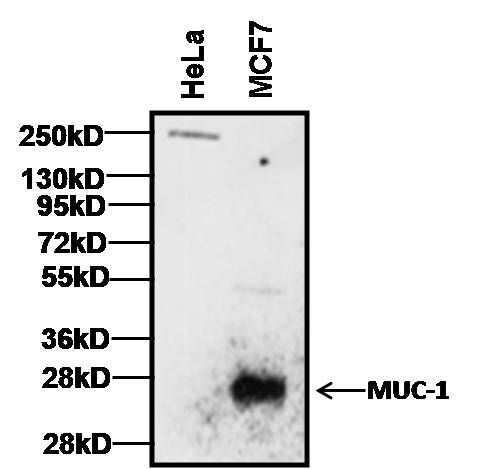
- Western blot analysis of MUC-1 (CT2) was performed by loading 25 µg of HeLa (left lane) and MCF7 (right lane) cell lysates, and 10 µL of PageRuler Plus Prestained Protein Ladder (Product # 26619) onto a Novex® 4-20% Tris-Glycine polyacrylamide gel. Proteins were transferred to a PVDF membrane using the G2 Fast Blotter (Product # 62288), and blocked with 5% milk in TBST for 1 hour at room temperature. Small subunits of MUC-1 were detected between ~14 kD to 30 kD mainly in MCF7 breast cancer cell samples using a MUC-1 monoclonal antibody (Product # MA5-11202) at a concentration of 1 µg/mL overnight at 4C on a rocking platform, followed by a goat anti-Armenian hamster IgG-HRP secondary antibody (Product # PA1-32045) at a dilution of 1:10,000 for at least 1 hour at room temperature. Chemiluminescent detection was performed using SuperSignal West Dura (Product # 34076).
- Submitted by
- Invitrogen Antibodies (provider)
- Main image
- Experimental details
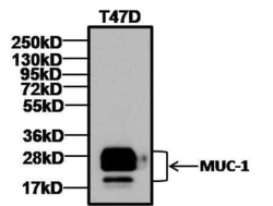
- Western blot analysis of MUC-1 (CT2) was performed by loading 25 µg of T47D cell lysates, and 10 µL of PageRuler Plus Prestained Protein Ladder (Product # 26619) onto a Novex® 4-20% Tris-Glycine polyacrylamide gel. Proteins were transferred to a PVDF membrane using the G2 Fast Blotter (Product # 62288), and blocked with 5% milk in TBST for 1 hour at room temperature. Small subunits of MUC-1 were detected between ~14 kD to 30 kD using a MUC-1 monoclonal antibody (Product # MA5-11202) at a concentration of 1 µg/mL overnight at 4C on a rocking platform, followed by a goat anti-Armenian hamster IgG-HRP secondary antibody (Product # PA1-32045) at a dilution of 1:10,000 for at least 1 hour at room temperature. Chemiluminescent detection was performed using SuperSignal West Dura (Product # 34076).

- Submitted by
- Invitrogen Antibodies (provider)
- Main image
- Experimental details
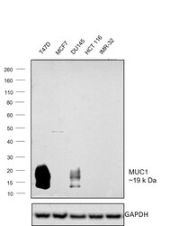
- Western blot was performed using Anti-MUC1 Monoclonal Antibody (MH1 (CT2)), (Product # MA5-11202) and ~19 kDa band corresponding to MUC1 was observed across cell lines tested except in MCF7, HCT 116 and IMR-32 which are reported to be negative. Whole cell extracts (30 µg lysate) of T47D (Lane 1), MCF7 (Lane 2), DU145 (Lane 3), HCT 116 (Lane 4) and IMR-32 (Lane 5) were electrophoresed using Novex® NuPAGE® 4-12 % Bis-Tris gel (Product # NP0321BOX). Resolved proteins were then transferred onto a nitrocellulose membrane (Product # IB23001) by iBlot® 2 Dry Blotting System (Product # IB21001). The blot was probed with the primary antibody (1µg/mL) and detected by chemiluminescence with Rabbit anti-Hamster IgG (H+L) Secondary Antibody, HRP (Product # A18889, 1:4000 dilution) using the iBright FL 1000 (Product # A32752). Chemiluminescent detection was performed using Super Signal™ West Dura Extended Duration Substrate (Product # 34076).

Supportive validation
- Submitted by
- Invitrogen Antibodies (provider)
- Main image
- Experimental details
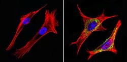
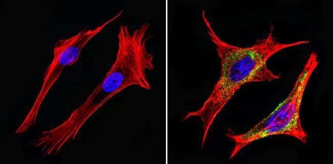
- Immunofluorescent analysis of CD227/MUC1/Mucin 1 (green) showing staining in the in the cytoplasm and membrane of NIH-3T3 cells (right) compared to a negative control without primary antibody (left). Formalin-fixed cells were permeabilized with 0.1% Triton X-100 in TBS for 5-10 minutes and blocked with 3% BSA-PBS for 30 minutes at room temperature. Cells were probed with a CD227/MUC1/Mucin 1 monoclonal antibody (Product # MA5-11202) in 3% BSA-PBS at a dilution of 1:50 and incubated overnight at 4ºC in a humidified chamber. Cells were washed with PBST and incubated with a DyLight-conjugated secondary antibody in PBS at room temperature in the dark. F-actin (red) was stained with a fluorescent red phalloidin and nuclei (blue) were stained with Hoechst or DAPI. Images were taken at a magnification of 60x.
- Submitted by
- Invitrogen Antibodies (provider)
- Main image
- Experimental details
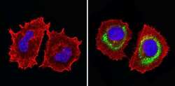
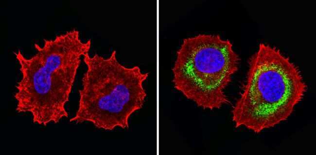
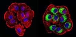
- Immunofluorescent analysis of CD227/MUC1/Mucin 1 (green) showing staining in the in the cytoplasm and membrane of MCF-7 cells (right) compared to a negative control without primary antibody (left). Formalin-fixed cells were permeabilized with 0.1% Triton X-100 in TBS for 5-10 minutes and blocked with 3% BSA-PBS for 30 minutes at room temperature. Cells were probed with a CD227/MUC1/Mucin 1 monoclonal antibody (Product # MA5-11202) in 3% BSA-PBS at a dilution of 1:50 and incubated overnight at 4ºC in a humidified chamber. Cells were washed with PBST and incubated with a DyLight-conjugated secondary antibody in PBS at room temperature in the dark. F-actin (red) was stained with a fluorescent red phalloidin and nuclei (blue) were stained with Hoechst or DAPI. Images were taken at a magnification of 60x.
- Submitted by
- Invitrogen Antibodies (provider)
- Main image

- Experimental details
- Immunofluorescent analysis of CD227/MUC1/Mucin 1 (green) showing staining in the in the cytoplasm and membrane of NIH-3T3 cells (right) compared to a negative control without primary antibody (left). Formalin-fixed cells were permeabilized with 0.1% Triton X-100 in TBS for 5-10 minutes and blocked with 3% BSA-PBS for 30 minutes at room temperature. Cells were probed with a CD227/MUC1/Mucin 1 monoclonal antibody (Product # MA5-11202) in 3% BSA-PBS at a dilution of 1:50 and incubated overnight at 4ºC in a humidified chamber. Cells were washed with PBST and incubated with a DyLight-conjugated secondary antibody in PBS at room temperature in the dark. F-actin (red) was stained with a fluorescent red phalloidin and nuclei (blue) were stained with Hoechst or DAPI. Images were taken at a magnification of 60x.
- Submitted by
- Invitrogen Antibodies (provider)
- Main image
- Experimental details
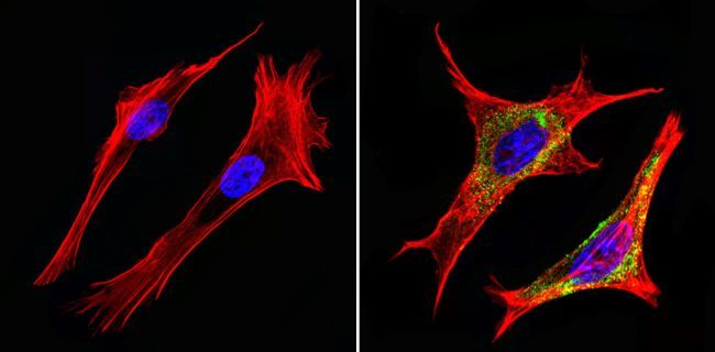
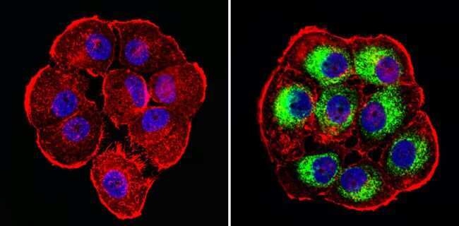
- Immunofluorescent analysis of CD227/MUC1/Mucin 1 (green) showing staining in the in the cytoplasm of T47D cells (right) compared to a negative control without primary antibody (left). Formalin-fixed cells were permeabilized with 0.1% Triton X-100 in TBS for 5-10 minutes and blocked with 3% BSA-PBS for 30 minutes at room temperature. Cells were probed with a CD227/MUC1/Mucin 1 monoclonal antibody (Product # MA5-11202) in 3% BSA-PBS at a dilution of 1:50 and incubated overnight at 4ºC in a humidified chamber. Cells were washed with PBST and incubated with a DyLight-conjugated secondary antibody in PBS at room temperature in the dark. F-actin (red) was stained with a fluorescent red phalloidin and nuclei (blue) were stained with Hoechst or DAPI. Images were taken at a magnification of 60x.
- Submitted by
- Invitrogen Antibodies (provider)
- Main image
- Experimental details
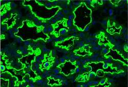
- Immunofluorescence analysis of MUC1 (green) in lactating mouse mammary tissue. FFPE sections were subjected to antigen retrieval at pH 6.0 followed by blocking with 1% BSA and 10% goat serum. Cells were stained with monoclonal MUC1 antibody (Product # MA5-11202) at a dilution of 1:50 at 4 deg overnight, followed by Armenian hamster 488 secondary antibody (green) and DAPI (blue) to stain the nuclei. Image courtesy of Antibody Data Exchange Program.

Supportive validation
- Submitted by
- Invitrogen Antibodies (provider)
- Main image

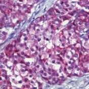
- Experimental details
- Formalin-fixed, paraffin-embedded human breast cancer stained with EMA antibody using alkaline phosphatase-conjugate and fast red chromogen. Note nuclear and cytoplasmic staining of tumor cells.
- Submitted by
- Invitrogen Antibodies (provider)
- Main image

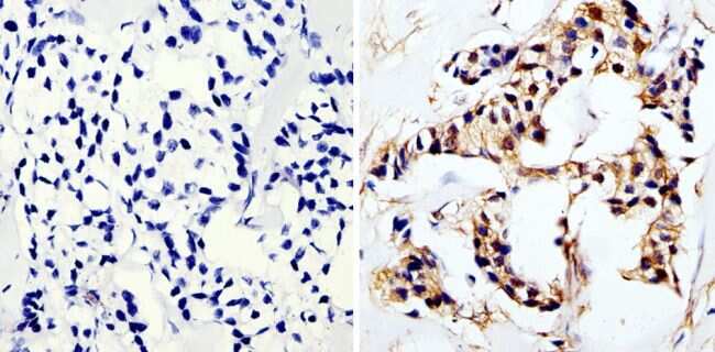
- Experimental details
- Immunohistochemistry analysis of Mucin 1 showing staining in the cytoplasm of paraffin-embedded human breast carcinoma (right) compared to a negative control without primary antibody (left). To expose target proteins, antigen retrieval was performed using 10mM sodium citrate (pH 6.0), microwaved for 8-15 min. Following antigen retrieval, tissues were blocked in 3% H2O2-methanol for 15 min at room temperature, washed with ddH2O and PBS, and then probed with a Mucin 1 monoclonal antibody (Product # MA5-11202) diluted in 3% BSA-PBS at a dilution of 1:20 overnight at 4°C in a humidified chamber. Tissues were washed extensively in PBST and detection was performed using an HRP-conjugated secondary antibody followed by colorimetric detection using a DAB kit. Tissues were counterstained with hematoxylin and dehydrated with ethanol and xylene to prep for mounting.
Supportive validation
- Submitted by
- Invitrogen Antibodies (provider)
- Main image
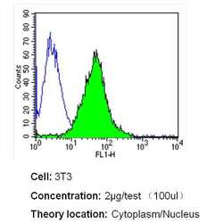
- Experimental details
- Flow cytometry analysis of Mucin 1 in NIH-3T3 cells (green) compared to an isotype control (blue). Cells were harvested, adjusted to a concentration of 1-5x10^6 cells/mL, fixed with 2% paraformaldehyde and washed with PBS. Cells were blocked with a 2% solution of BSA-PBS for 30 min at room temperature and incubated with a Mucin 1 monoclonal antibody (Product # MA5-11202) at a dilution of 2 µg/test for 40 min at room temperature. Cells were then incubated for 40 min at room temperature in the dark using a Dylight 488-conjugated secondary antibody and re-suspended in PBS for FACS analysis.

- Submitted by
- Invitrogen Antibodies (provider)
- Main image

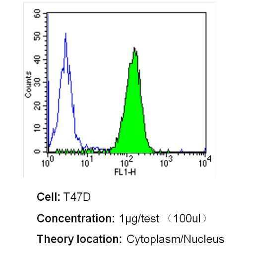
- Experimental details
- Flow cytometry analysis of Mucin 1 in T47D cells (green) compared to an isotype control (blue). Cells were harvested, adjusted to a concentration of 1-5x10^6 cells/mL, fixed with 2% paraformaldehyde and washed with PBS. Cells were blocked with a 2% solution of BSA-PBS for 30 min at room temperature and incubated with a Mucin 1 monoclonal antibody (Product # MA5-11202) at a dilution of 1 µg/test for 40 min at room temperature. Cells were then incubated for 40 min at room temperature in the dark using a Dylight 488-conjugated secondary antibody and re-suspended in PBS for FACS analysis.
- Submitted by
- Invitrogen Antibodies (provider)
- Main image
- Experimental details
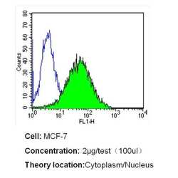
- Flow cytometry analysis of Mucin 1 in MCF-7 cells (green) compared to an isotype control (blue). Cells were harvested, adjusted to a concentration of 1-5x10^6 cells/mL, fixed with 2% paraformaldehyde and washed with PBS. Cells were blocked with a 2% solution of BSA-PBS for 30 min at room temperature and incubated with a Mucin 1 monoclonal antibody (Product # MA5-11202) at a dilution of 2 µg/test for 40 min at room temperature. Cells were then incubated for 40 min at room temperature in the dark using a Dylight 488-conjugated secondary antibody and re-suspended in PBS for FACS analysis.

Supportive validation
- Submitted by
- Invitrogen Antibodies (provider)
- Main image

- Experimental details
- NULL
- Submitted by
- Invitrogen Antibodies (provider)
- Main image
- Experimental details
- NULL

- Submitted by
- Invitrogen Antibodies (provider)
- Main image
- Experimental details
- NULL
- Submitted by
- Invitrogen Antibodies (provider)
- Main image
- Experimental details
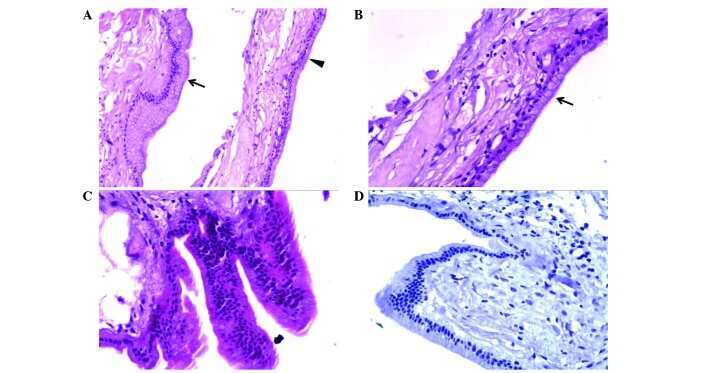
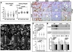
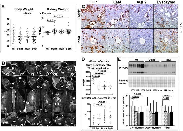
- Fig 3 A) Body weight and kidney weight as percent of body weight (%KW/BW) in wild-type mice and in Pde1a Del15 and Pde1a InsA homozygous mice or data from both combined. B) Axial and coronal MR images of Pde1a mutant mice at 6 and 12 months of age showing small renal cysts (arrows; this panel is also shown as a larger figure in Fig B in S1 File ). C) Kidney sections from Pde1a Del15 and Pde1a InsA homozygous mice showing multiple small cysts staining positively for Tamm-Horsfall protein and epithelial membrane antigen, less consistently for aquaporin-2, and not staining for lysozyme. D) Reduced urine osmolality after 24 hours of dehydration, faster excretion of an acute water load, and E) reduced expression of glycosylated (upper band) and unglycosylated (lower band) pSer269-AQP2 in Pde1a Del15 and Pde1a InsA homozygotes compared to wild-type mice. One-way ANOVA with post-hoc Tukey test was used for the statistical analysis.
- Submitted by
- Invitrogen Antibodies (provider)
- Main image

- Experimental details
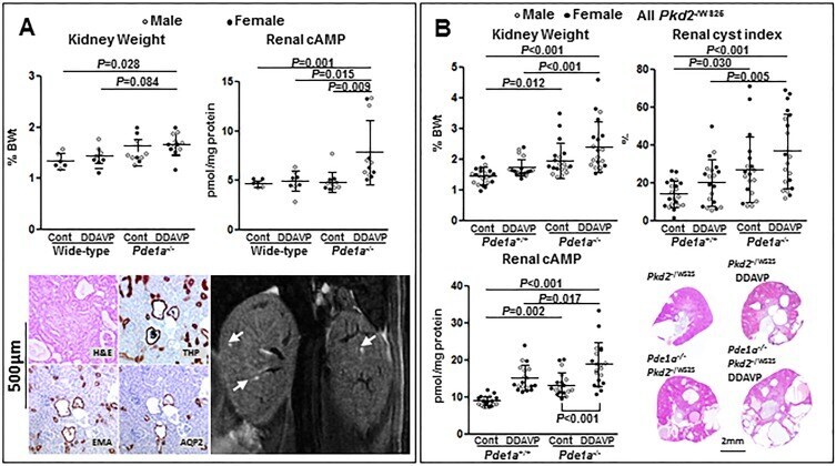
- Fig 5 A) Upper panels: Kidney weights as percent of body weights (%KW/BW) and renal cAMP levels in untreated or desmopressin treated wild-type and Pde1a InsA homozygous mice (upper panels); One-way ANOVA with post-hoc Tukey test was used for the statistical analysis; using a two-way ANOVA, both Pde1a genotype P
- Submitted by
- Invitrogen Antibodies (provider)
- Main image

- Experimental details
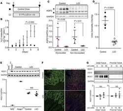
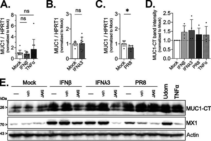
- FIG 3 Cell-associated MUC1 levels are upregulated during IAV infection and IFN treatment. HAE were stimulated with (A) IFN-beta, (A) TNF-alpha, or (B) IFN-lambda3 or (C) infected with PR8 (5 x 10 4 PFU; approximate MOI of 1), and MUC1 expression was quantified by qPCR after 24 h of treatment. (D) HAE were stimulated as indicated or infected with PR8 as for panels A to C for 24 h, protein lysate collected, and MUC1 expression quantified by Western blotting for MUC1-CT. MUC1-CT band intensity was analyzed by densitometry relative to actin band intensity. (E) HAE were stimulated with IFN-beta, IFN-lambda3, or PR8 alone (-) or in the presence of the JAK inhibitor ruxolitinib (JAKi) or DMSO as a vehicle control (veh). Additional cultures were stimulated with Udorn or TNF-alpha alone. After 24 h, lysate was collected and analyzed by Western blotting for MUC1-CT, MX1, or actin. Results in panels A to C are from three experimental replicates utilizing three different HAE donors with a minimum of three biological replicates from each donor. The densitometry analysis (D) shows data from four experimental replicates utilizing four different HAE donors with one biological replicate from each donor. All experimental results were analyzed by the Mann-Whitney U test compared to mock conditions and are significant where indicated (*, P < 0.05; ns, not significant).
- Submitted by
- Invitrogen Antibodies (provider)
- Main image

- Experimental details
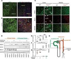
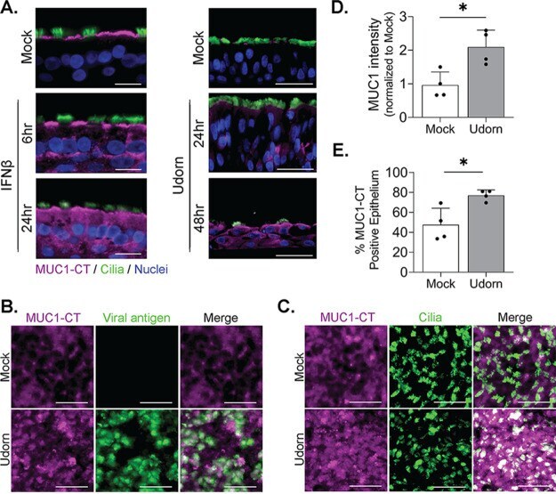
- FIG 4 Type 1 IFN and IAV broadly upregulate MUC1 expression across HAE. (A) HAE were stimulated with IFN-beta or mock stimulated (left) or infected with Udorn (5 x 10 2 PFU, approximate MOI of 0.01) (right) and fixed for immunohistochemical detection of MUC1-CT (purple), acetylated alpha-tubulin (cilium marker; green), and nuclei at the indicated time points. (B to E) HAE were infected with Udorn (5 x 10 4 PFU, approximate MOI of 1), fixed at 24 hpi, and stained en face for MUC1-CT (purple) along with (B) viral antigen (nucleoprotein, green), or (C) a ciliated cell marker (acetylated alpha-tubulin; green). The mean intensity (D) or total area staining positive for MUC1-CT (E) was quantified by FIJI on four additional cultures across two donors after infection as for panels B and C and analyzed by the Mann-Whitney U test compared to the mock condition, indicating significance (*, P < 0.05). Results in panel A are from one experimental replicate utilizing two different HAE donors (left and right) and one biological replicate from each donor. Results in panels B and C are from the same donor. Results in panels D and E are from two experimental replicates utilizing two different HAE donors with two biological replicates from each donor. Bars = 20 mum (A) and 25 mum (B and C).
- Submitted by
- Invitrogen Antibodies (provider)
- Main image

- Experimental details
- FIG 6 Establishment and characterization of immortalized HAE depleted of MUC1. Immortalized airway epithelial BCi-NS1.1 cells were transduced with CRISPR/Cas9 and sgRNA targeting (A) MUC1 exon 5 for protein depletion (MUC1 KO ) or without (control [Ctl]) the predicted targeting site. (B) Genomic DNA was extracted and used in a T7 endonuclease I cleavage assay demonstrating editing at the target site. (C) After differentiation, total HAE lysate was collected, separated by PAGE, and blotted for nontargeted tethered mucin MUC4 (extracellular domain), MUC1-ED, MUC1-CT, and actin. (D) Representative histological sections of paraffin-embedded cultures show normal ciliated epithelium. H&E counterstain. Bar = 20 mum. (E) Fluorescent microparticles were applied apically to indicated cultures to determine mucociliary transport rate. MCC between culture types was analyzed by the Mann-Whitney U test (****, P < 0.0001).

- Submitted by
- Invitrogen Antibodies (provider)
- Main image
- Experimental details
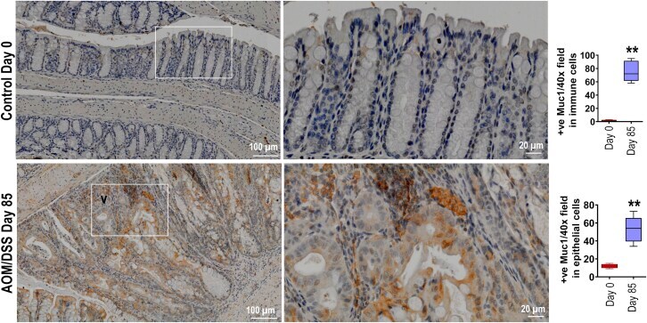
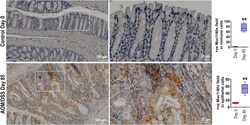
- Figure 12 MUC1 is upregulated in both tumor and immune cells in repose to AOM/DSS. MUC1 IHC staining colon section from WT mice ether treated with AOM followed by DSS as per Figure 1 , A at day 85 or untreated at day 0 (n >= 6). Statistics: box plots show median, quartiles and range. Mann-Whitney U test. * vs Day 0, ** P < .01. The data are representative of 3 independent experiments.