Explore
 Validate
 Learn
LearnGTX25621
antibody from GeneTex
Targeting: THRA
AR7, EAR-7.1/EAR-7.2, ERBA, ERBA1, NR1A1, THRA1, THRA2, THRA3
 Western blot
 Immunohistochemistry
Antibody data
- Antibody Data
- Antigen structure
- References [2]
- Comments [0]
- Validations
- Immunohistochemistry [3]
Submit
Validation data
Reference
Comment
Report error
- Product number
- GTX25621 - Provider product page

- Provider
- GeneTex
- Proper citation
- GeneTex Cat#GTX25621, RRID:AB_380602
- Product name
- Thyroid Hormone Receptor alpha antibody
- Antibody type
- Polyclonal
- Reactivity
- Human, Mouse, Rat, Chicken/Avian, Sheep, Simian, Xenopus, Zebrafish
- Host
- Rabbit
Submitted references Sex Differences in Brain Thyroid Hormone Levels during Early Post-Hatching Development in Zebra Finch (Taeniopygia guttata).
Thyroid hormone suppresses expression of stathmin and associated tumor growth in hepatocellular carcinoma.
Yamaguchi S, Hayase S, Aoki N, Takehara A, Ishigohoka J, Matsushima T, Wada K, Homma KJ
PloS one 2017;12(1):e0169643
PloS one 2017;12(1):e0169643
Thyroid hormone suppresses expression of stathmin and associated tumor growth in hepatocellular carcinoma.
Tseng YH, Huang YH, Lin TK, Wu SM, Chi HC, Tsai CY, Tsai MM, Lin YH, Chang WC, Chang YT, Chen WJ, Lin KH
Scientific reports 2016 Dec 9;6:38756
Scientific reports 2016 Dec 9;6:38756
No comments: Submit comment
Supportive validation
- Submitted by
- GeneTex (provider)
- Main image

- Experimental details
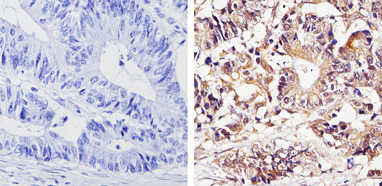
- Immunohistochemistry analysis of Thyroid Hormone Receptor alpha-1 showing staining in the cytoplasm and nucleus of paraffin-treated human colon carcinoma (right) compared with a negative control in the absence of primary antibody (left). To expose target proteins, antigen retrieval was performed using 10mM sodium citrate (pH 6.0), microwaved for 8-15 min. Following antigen retrieval, tissues were blocked in 3% H2O2-methanol for 15 min at room temperature, washed with ddH2O and PBS, and then probed with a Thyroid Hormone Receptor alpha-1 polyclonal antibody (GTX25621) diluted by 3% BSA-PBS at a dilution of 1:500 overnight at 4¢XC in a humidified chamber. Tissues were washed extensively in PBST and detection was performed using an HRP-conjugated secondary antibody followed by colorimetric detection using a DAB kit. Tissues were counterstained with hematoxylin and dehydrated with ethanol and xylene to prep for mounting.
- Submitted by
- GeneTex (provider)
- Main image

- Experimental details
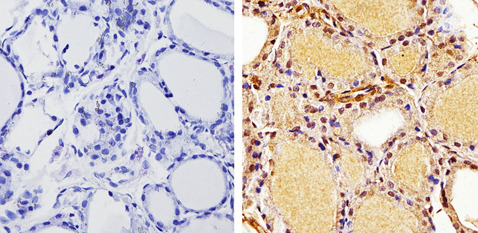
- Immunohistochemistry analysis of Thyroid Hormone Receptor alpha-1 showing staining in the cytoplasm and nucleus of paraffin-treated human thyroid tissue (right) compared with a negative control in the absence of primary antibody (left). To expose target proteins, antigen retrieval was performed using 10mM sodium citrate (pH 6.0), microwaved for 8-15 min. Following antigen retrieval, tissues were blocked in 3% H2O2-methanol for 15 min at room temperature, washed with ddH2O and PBS, and then probed with a Thyroid Hormone Receptor alpha-1 polyclonal antibody (GTX25621) diluted by 3% BSA-PBS at a dilution of 1:500 overnight at 4¢XC in a humidified chamber. Tissues were washed extensively in PBST and detection was performed using an HRP-conjugated secondary antibody followed by colorimetric detection using a DAB kit. Tissues were counterstained with hematoxylin and dehydrated with ethanol and xylene to prep for mounting.
- Submitted by
- GeneTex (provider)
- Main image
- Experimental details
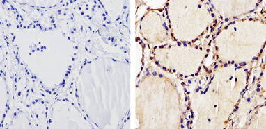
- Immunohistochemistry analysis of Thyroid Hormone Receptor alpha-1 showing staining in the cytoplasm and nucleus of paraffin-treated rat thyroid tissue (right) compared with a negative control in the absence of primary antibody (left). To expose target proteins, antigen retrieval was performed using 10mM sodium citrate (pH 6.0), microwaved for 8-15 min. Following antigen retrieval, tissues were blocked in 3% H2O2-methanol for 15 min at room temperature, washed with ddH2O and PBS, and then probed with a Thyroid Hormone Receptor alpha-1 polyclonal antibody (GTX25621) diluted by 3% BSA-PBS at a dilution of 1:200 overnight at 4¢XC in a humidified chamber. Tissues were washed extensively in PBST and detection was performed using an HRP-conjugated secondary antibody followed by colorimetric detection using a DAB kit. Tissues were counterstained with hematoxylin and dehydrated with ethanol and xylene to prep for mounting.