Explore
 Validate
 Learn
Learn Western blot
Antibody data
- Antibody Data
- Antigen structure
- References [1]
- Comments [0]
- Validations
- Western blot [2]
- Immunohistochemistry [7]
Submit
Validation data
Reference
Comment
Report error
- Product number
- NBP1-92012 - Provider product page

- Provider
- Novus Biologicals
- Proper citation
- Novus Cat#NBP1-92012, RRID:AB_11016139
- Product name
- Rabbit Polyclonal HspA1L Antibody
- Antibody type
- Polyclonal
- Description
- Immunogen affinity purified. Specificity of human, mouse, rat HspA1L antibody verified on a Protein Array containing target protein plus 383 other non-specific proteins.
- Reactivity
- Human, Mouse, Rat
- Host
- Rabbit
- Isotype
- IgG
- Vial size
- 0.1 ml
- Storage
- Store at 4C short term. Aliquot and store at -20C long term. Avoid freeze-thaw cycles.
Submitted references Melatonin Enhances Mitophagy by Upregulating Expression of Heat Shock 70 kDa Protein 1L in Human Mesenchymal Stem Cells under Oxidative Stress.
Yoon YM, Kim HJ, Lee JH, Lee SH
International journal of molecular sciences 2019 Sep 13;20(18)
International journal of molecular sciences 2019 Sep 13;20(18)
No comments: Submit comment
Supportive validation
- Submitted by
- Novus Biologicals (provider)
- Main image

- Experimental details
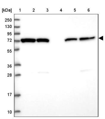
- Western Blot: HspA1L Antibody [NBP1-92012] - Lane 1: Marker [kDa] 250, 130, 95, 72, 55, 36, 28, 17, 10. Lane 2: Human cell line RT-4. Lane 3: Human cell line U-251MG sp. Lane 4: Human plasma (IgG/HSA depleted). Lane 5: Human liver tissue. Lane 6: Human tonsil tissue
- Submitted by
- Novus Biologicals (provider)
- Main image
- Experimental details
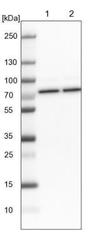
- Western Blot: HspA1L Antibody [NBP1-92012] - Lane 1: NIH-3T3 cell lysate (Mouse embryonic fibroblast cells). Lane 2: NBT-II cell lysate (Rat Wistar bladder tumor cells).

Supportive validation
- Submitted by
- Novus Biologicals (provider)
- Main image

- Experimental details
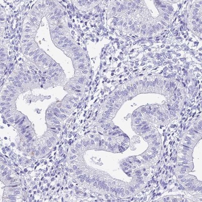
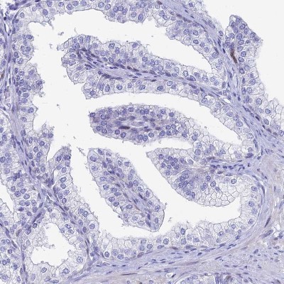
- Immunohistochemistry-Paraffin: HspA1L Antibody [NBP1-92012] - Staining of human endometrium shows low expression as expected.
- Submitted by
- Novus Biologicals (provider)
- Main image

- Experimental details
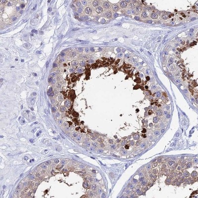
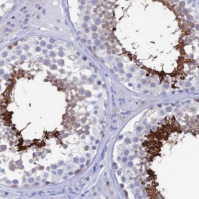
- Immunohistochemistry-Paraffin: HspA1L Antibody [NBP1-92012] - Staining of human testis shows high expression.
- Submitted by
- Novus Biologicals (provider)
- Main image
- Experimental details
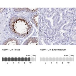
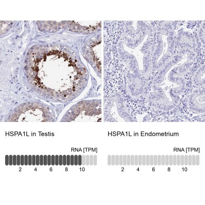
- Immunohistochemistry-Paraffin: HspA1L Antibody [NBP1-92012] - Analysis in human testis and endometrium tissues. Corresponding HspA1L RNA-seq data are presented for the same tissues.
- Submitted by
- Novus Biologicals (provider)
- Main image

- Experimental details
- Immunohistochemistry-Paraffin: HspA1L Antibody [NBP1-92012] - Staining of human endometrium shows no positivity in glandular cells as expected.

- Submitted by
- Novus Biologicals (provider)
- Main image

- Experimental details
- Immunohistochemistry-Paraffin: HspA1L Antibody [NBP1-92012] - Staining of human kidney shows no positivity in cells in tubules as expected.

- Submitted by
- Novus Biologicals (provider)
- Main image

- Experimental details
- Immunohistochemistry-Paraffin: HspA1L Antibody [NBP1-92012] - Staining of human prostate shows no positivity in glandular cells as expected.
- Submitted by
- Novus Biologicals (provider)
- Main image
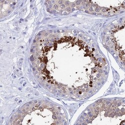
- Experimental details
- Immunohistochemistry-Paraffin: HspA1L Antibody [NBP1-92012] - Staining of human testis shows strong cytoplasmic positivity in cells in seminiferous ducts.