Explore
 Validate
 Learn
Learn Western blot
Antibody data
- Antibody Data
- Antigen structure
- References [0]
- Comments [0]
- Validations
- Western blot [2]
- Immunocytochemistry [2]
Submit
Validation data
Reference
Comment
Report error
- Product number
- 703632 - Provider product page

- Provider
- Invitrogen Antibodies
- Product name
- SLIRP Recombinant Rabbit Monoclonal Antibody (3H17L4)
- Antibody type
- Monoclonal
- Antigen
- Other
- Description
- This antibody is predicted to react with Monkey, Horse, Cat, Bovine.
- Antibody clone number
- 3H17L4
- Concentration
- 0.5 mg/mL
No comments: Submit comment
Supportive validation
- Submitted by
- Invitrogen Antibodies (provider)
- Main image

- Experimental details
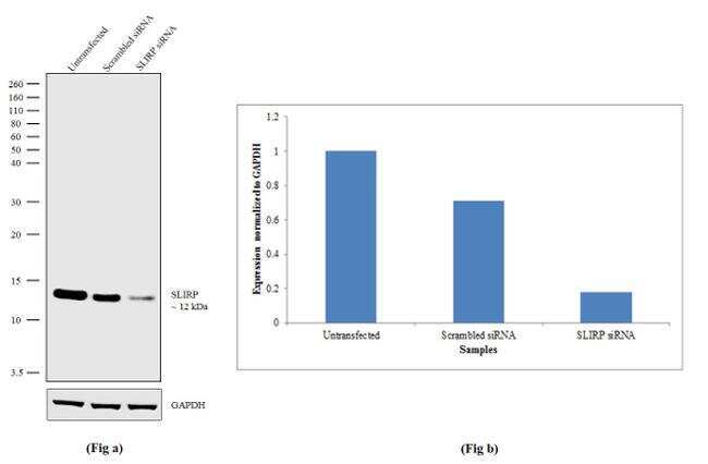
- Knockdown of SLIRP was achieved by transfecting MCF7 cells with SLIRP specific siRNA (Silencer® select Product # s37869 & s37870). Western blot analysis (Fig a) was performed using modified whole cell extracts (1% SDS) from SLIRP knockdown cells (Lane 3), non-specific scrambled siRNA transfected cells (Lane 2) and untransfected cells (Lane 1). The blot was probed with Anti-SLIRP Recombinant Rabbit Monoclonal Antibody (Product # 703632, 1:5000 dilution) and Goat anti-Rabbit IgG (H+L) Superclonal™ Secondary Antibody, HRP conjugate (Product # A27036, 0.25 µg/mL, 1:4000 dilution). Densitometric analysis of this Western blot is shown in histogram (Fig b). Loss of signal upon siRNA mediated knockdown confirms that antibody is specific to SLIRP.
- Submitted by
- Invitrogen Antibodies (provider)
- Main image

- Experimental details
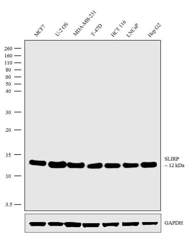
- Western blot analysis was performed on modified whole cell extracts (1% SDS) (30 µg lysate) of MCF7 (Lane 1), U-2 OS (Lane 2), MDA-MB-231 (Lane 3), T-47D (Lane 4), HCT 116 (Lane 5), LNCaP (Lane 6) and Hep G2 (Lane 7). The blot was probed with Anti-SLIRP Recombinant Rabbit Monoclonal Antibody (Product # 703632, 1:5000 dilution) and detected by chemiluminescence using Goat anti-Rabbit IgG (H+L) Superclonal™ Secondary Antibody, HRP conjugate (Product # A27036, 0.25 µg/mL, 1:4000 dilution). A ~12 kDa band corresponding to SLIRP was observed across the cell lines tested.
Supportive validation
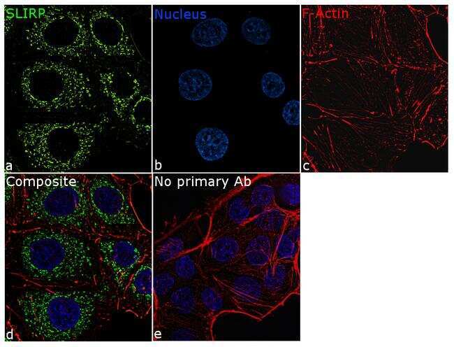
- Submitted by
- Invitrogen Antibodies (provider)
- Main image

- Experimental details
- For immunofluorescence analysis, MCF7 cells were fixed and permeabilized for detection of endogenous SLIRP using Anti-SLIRP Recombinant Rabbit Monoclonal Antibody (Product # 703632, 1:100 dilution) and labeled with Goat anti-Rabbit IgG (H+L) Superclonal™ Secondary Antibody, Alexa Fluor® 488 conjugate (Product # A27034, 1:2000). Panel a) shows representative cells that were stained for detection and localization of SLIRP protein (green), Panel b) is stained for nuclei (blue) using ProLong™ Diamond Antifade Mountant with DAPI (Product # P36962). Panel c) represents cytoskeletal F-actin staining using Rhodamine Phalloidin (Product # R415, 1:300). Panel d) is a composite image of Panels a, b and c clearly demonstrating mitochondrial localization of SLIRP. Panel e) represents control cells with no primary antibody to assess background. The images were captured at 60X magnification.
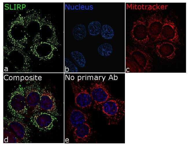
- Submitted by
- Invitrogen Antibodies (provider)
- Main image
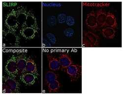
- Experimental details
- For immunofluorescence analysis, MCF7 cells were fixed and permeabilized for detection of endogenous SLIRP using Anti-Citrate synthase Recombinant Rabbit Monoclonal Antibody (Product # 703632, 1:100 dilution) and labeled with Goat anti-Rabbit IgG (H+L) Superclonal™ Secondary Antibody, Alexa Fluor® 488 conjugate (Product # A27034, 1:2000). Panel a) shows representative cells that were stained for detection and localization of SLIRP protein (green), Panel b) is stained for nuclei (blue) using ProLong™ Diamond Antifade Mountant with DAPI (Product # P36962). Panel c) represents mitochondrial staining using MitoTracker® Red CMXRos (Product # M7512). Panel d) is a composite image of Panels a, b and c clearly demonstrating co-localization of SLIRP with mitotracker which specifically binds to the mitochondria. Panel e) represents control cells without primary antibody to assess background. The images were captured at 60X magnification.