Explore
 Validate
 Learn
Learn Western blot
Antibody data
- Antibody Data
- Antigen structure
- References [3]
- Comments [0]
- Validations
- Western blot [7]
- Immunocytochemistry [2]
- Immunohistochemistry [3]
Submit
Validation data
Reference
Comment
Report error
- Product number
- GTX103219 - Provider product page

- Provider
- GeneTex
- Proper citation
- GeneTex Cat#GTX103219, RRID:AB_1949578
- Product name
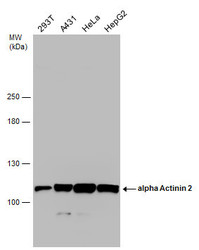
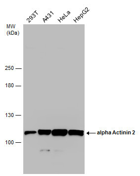
- alpha Actinin 2 antibody [N1N3]
- Antibody type
- Polyclonal
- Reactivity
- Human, Mouse, Rat, Porcine
- Host
- Rabbit
Submitted references The effects of artocarpin on wound healing: in vitro and in vivo studies.
ATF6 Decreases Myocardial Ischemia/Reperfusion Damage and Links ER Stress and Oxidative Stress Signaling Pathways in the Heart.
Cardiac Depression in Pigs after Multiple Trauma - Characterization of Posttraumatic Structural and Functional Alterations.
Yeh CJ, Chen CC, Leu YL, Lin MW, Chiu MM, Wang SH
Scientific reports 2017 Nov 15;7(1):15599
Scientific reports 2017 Nov 15;7(1):15599
ATF6 Decreases Myocardial Ischemia/Reperfusion Damage and Links ER Stress and Oxidative Stress Signaling Pathways in the Heart.
Jin JK, Blackwood EA, Azizi K, Thuerauf DJ, Fahem AG, Hofmann C, Kaufman RJ, Doroudgar S, Glembotski CC
Circulation research 2017 Mar 3;120(5):862-875
Circulation research 2017 Mar 3;120(5):862-875
Cardiac Depression in Pigs after Multiple Trauma - Characterization of Posttraumatic Structural and Functional Alterations.
Kalbitz M, Schwarz S, Weber B, Bosch B, Pressmar J, Hoenes FM, Braun CK, Horst K, Simon TP, Pfeifer R, Störmann P, Hummler H, Gebhard F, Pape HC, Huber-Lang M, Hildebrand F, TREAT Research Group.
Scientific reports 2017 Dec 19;7(1):17861
Scientific reports 2017 Dec 19;7(1):17861
No comments: Submit comment
Supportive validation
- Submitted by
- GeneTex (provider)
- Main image

- Experimental details
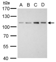
- Sample(30 ug whole cell lysate)A:A431(GTX27909)B:H1299C:HeLa S3(GTX14654)D:Hep G2 (GTX27900)7.5% SDS PAGEGTX103219 diluted at 1:1000
- Validation comment
- WB
- Submitted by
- GeneTex (provider)
- Main image
- Experimental details
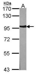
- Sample (30 ?g of whole cell lysate) A:NIH-3T37.5% SDS PAGE GTX103219 diluted at 1:1000 The HRP-conjugated anti-rabbit IgG antibody (GTX213110-01) was used to detect the primary antibody.

- Submitted by
- GeneTex (provider)
- Main image
- Experimental details
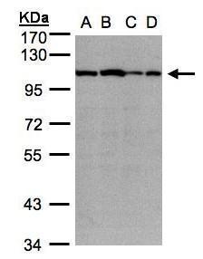
- Alpha Actinin 2 antibody [N1N3] detects ACTN2 protein by western blot analysis.A. 30 ?g A431 whole cell lysate/extractB. 30 ?g HeLa whole cell lysate/extractC. 30 ?g HepG2 whole cell lysate/extractD. 30 ?g A375 whole cell lysate/extract7.5 % SDS-PAGEalpha Actinin 2 antibody [N1N3] (GTX103219) dilution: 1:1000
- Validation comment
- WB

- Submitted by
- GeneTex (provider)
- Main image
- Experimental details
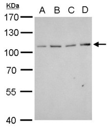
- Alpha Actinin 2 antibody [N1N3] detects ACTN2 protein by western blot analysis.A. 30 ?g Neuro2A whole cell lysate/extractB. 30 ?g GL261 whole cell lysate/extractC. 30 ?g C8D30 whole cell lysate/extractD. 30 ?g NIH-3T3 whole cell lysate/extract7.5% SDS-PAGEalpha Actinin 2 antibody [N1N3] (GTX103219) dilution: 1:1000The HRP-conjugated anti-rabbit IgG antibody (GTX213110-01) was used to detect the primary antibody.

- Submitted by
- GeneTex (provider)
- Main image
- Experimental details
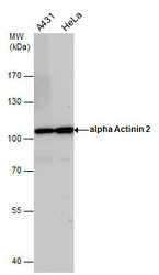
- alpha Actinin 2 antibody detects alpha Actinin 2 protein by western blot analysis. Various whole cell extracts (30 £gg) were separated by 7.5% SDS-PAGE, and the membrane was blotted with alpha Actinin 2 antibody (GTX103219) diluted by 1:1000.
- Submitted by
- GeneTex (provider)
- Main image
- Experimental details
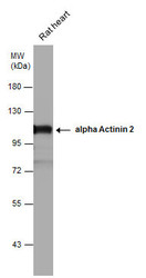
- alpha Actinin 2 antibody detects alpha Actinin 2 protein by western blot analysis. Rat tissue extracts (50 ?g) was separated by 7.5% SDS-PAGE, and the membrane was blotted with alpha Actinin 2 antibody (GTX103219) diluted at 1:5000. The HRP-conjugated anti-rabbit IgG antibody (GTX213110-01) was used to detect the primary antibody.

- Submitted by
- GeneTex (provider)
- Main image
- Experimental details
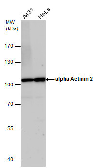
- Various whole cell extracts (30 ?g) were separated by 5% SDS-PAGE, and the membrane was blotted with alpha Actinin 2 antibody [N1N3] (GTX103219) diluted at 1:1000. The HRP-conjugated anti-rabbit IgG antibody (GTX213110-01) was used to detect the primary antibody.
Supportive validation
- Submitted by
- GeneTex (provider)
- Main image
- Experimental details
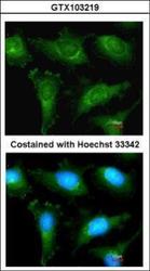
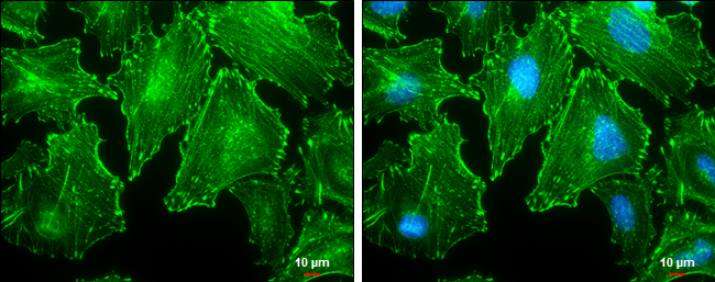
- Immunofluorescence analysis of methanol-fixed HeLa, using alpha Actinin 2(GTX103219) antibody at 1:200 dilution.

- Submitted by
- GeneTex (provider)
- Main image

- Experimental details
- alpha Actinin 2 antibody [N1N3] detects alpha Actinin 2 protein at cytoskeleton by immunofluorescent analysis.Sample: HeLa cells were fixed in ice-cold MeOH for 5 min.Green: alpha Actinin 2 protein stained by alpha Actinin 2 antibody [N1N3] (GTX103219) diluted at 1:500.Blue: Hoechst 33342 staining.Scale bar = 10 £gm.
Supportive validation
- Submitted by
- GeneTex (provider)
- Main image
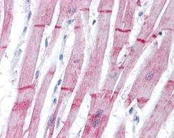
- Experimental details
- Immunohistochemical analysis of paraffin-embedded human heart, using alpha Actinin 2(GTX103219) antibody(10 £gg/ml).

- Submitted by
- GeneTex (provider)
- Main image

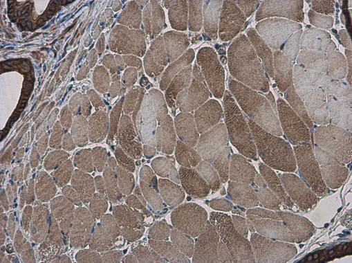
- Experimental details
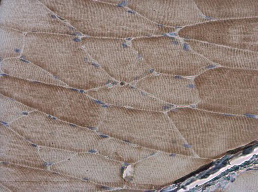
- alpha Actinin 2 antibody [N1N3] detects alpha Actinin 2 protein at cytoplasm in mouse muscle by immunohistochemical analysis. Sample: Paraffin-embedded mouse muscle. alpha Actinin 2 antibody [N1N3] (GTX103219) diluted at 1:500.
- Submitted by
- GeneTex (provider)
- Main image

- Experimental details
- alpha Actinin 2 antibody [N1N3] detects alpha Actinin 2 protein at cytoplasm in rat muscle by immunohistochemical analysis. Sample: Paraffin-embedded rat muscle. alpha Actinin 2 antibody [N1N3] (GTX103219) diluted at 1:500.