 Explore
 Validate
 Learn
Learn Western blot
 Immunohistochemistry
Antibody data
- Antibody Data
- Antigen structure
- References [0]
- Comments [0]
- Validations
- Western blot [2]
- Immunocytochemistry [2]
Submit
Validation data
Reference
Comment
Report error
- Product number
- PA1-46137 - Provider product page

- Provider
- Invitrogen Antibodies
- Product name
- GLUT10 Polyclonal Antibody
- Antibody type
- Polyclonal
- Antigen
- Other
- Reactivity
- Human, Mouse
- Host
- Rabbit
- Isotype
- IgG
- Vial size
- 100 µg
- Concentration
- 1 mg/mL
- Storage
- Store at 4°C short term. For long term storage, store at -20°C, avoiding freeze/thaw cycles.
No comments: Submit comment
Supportive validation
- Submitted by
- Invitrogen Antibodies (provider)
- Main image
- Experimental details
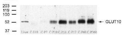
- Western blot analysis of GLUT10 in cochlea from embryonic mice and mice up to 1 yr old. Samples were incubated in GLUT10 polyclonal antibody (Product # PA1-46137) using a dilution of 1:500 followed by secondary at a dilution of 1:3,000.

- Submitted by
- Invitrogen Antibodies (provider)
- Main image

- Experimental details
- Western blot analysis of GLUT10 in NCI-H1299 non-small cell lung tumor cells. Samples were incubated in GLUT10 polyclonal antibody (Product # PA1-46137). Cells were transfected with the eukaryotic expression vectors pcDNA3.1/V5 (lanes 1,2), pCMV-GLUT10 (lanes 3,4), and pCMV-GLUT1 (lanes 5,6)..

Supportive validation
- Submitted by
- Invitrogen Antibodies (provider)
- Main image

- Experimental details
- Immunocytochemistry analysis of GLUT10 in dissected inner ear tissues from mice immersed in 20% sucrose PBS solution (pH 7.4) followed by being embedded in OCT. Samples were incubated in GLUT10 polyclonal antibody (Product # PA1-46137) using a dilution of 1:200 at 4 °C followed by anti-rabbit Cy3 secondary antibody with a dilution of 1:400 at room temperature for 1 hour. Tissues snap frozen in liquid nitrogen & 7 µm sections were cut by cryostat. Cochlear sections from organ of Corti were dissected & rinsed by 0.1% Triton in PBS (pH 7.4, PBST) for 30 min. Sections were blocked with 5% goat serum in PBST at RT 1 hr. Slides mounted with fluoromont G and examined under a confocal microscope. Strong immunoreactivity (red) is observed in the inner and outer hair cells and basilar membrane. Spiral ganglion neurons display moderate staining.
