 Explore
 Validate
 Learn
LearnSP5211P
antibody from Acris Antibodies GmbH
Targeting: NR3C1
GR, GRL
 Western blot Immunocytochemistry
 Immunoprecipitation Immunohistochemistry Immunoelectron microscopy Gel shift
Antibody data
- Antibody Data
- Antigen structure
- References [0]
- Comments [0]
- Validations
- Western blot [1]
- Immunocytochemistry [3]
- Immunohistochemistry [1]
Submit
Validation data
Reference
Comment
Report error
- Product number
- SP5211P - Provider product page

- Provider
- Acris Antibodies GmbH
- Proper citation
- Acris Antibodies GmbH Cat#SP5211P, RRID:AB_1001830
- Product name
- anti Glucocorticoid receptor
- Antibody type
- Polyclonal
- Antigen
- Synthetic Peptide corresponding to amino acid residues 150-175 from Human GR.
- Reactivity
- Human, Rat
- Host
- Rabbit
- Vial size
- 0.2 ml
No comments: Submit comment
Supportive validation
- Submitted by
- Acris Antibodies GmbH (provider)
- Main image
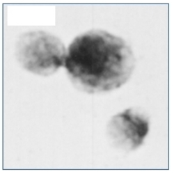
- Experimental details
- Immunolocalization of GR in human lymphocytes using Glucocorticoid Receptor antibody Cat.-No SP5211P.

Supportive validation
- Submitted by
- Acris Antibodies GmbH (provider)
- Main image
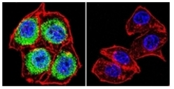
- Experimental details
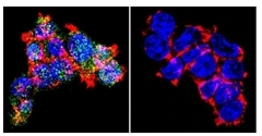
- Immunofluorescent analysis of Glucocorticoid Receptor using Glucocorticoid Receptor Polyclonal Antibody Cat.-No SP5211P shows staining in  HeLa Cells. Glucocorticoid Receptor (green), F-Actin staining with Phalloidin (red) and nuclei with DAPI (blue) is shown. Cells were grown on chamber slides and fixed with formaldehyde prior to staining. Cells were probed without (control) or with an antibody recognizing Glucocorticoid Receptor Cat.-No SP5211P at a dilution of 1/100 over night at 4°C, washed with PBS and incubated with a DyLight-488 conjugated secondary antibody. Images wee taken at 60x magnification.
- Submitted by
- Acris Antibodies GmbH (provider)
- Main image

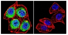
- Experimental details
- Immunofluorescent analysis of Glucocorticoid Receptor using Glucocorticoid Receptor Polyclonal Antibody Cat.-No SP5211P shows staining in  A2058 Cells. Glucocorticoid Receptor (green), F-Actin staining with Phalloidin (red) and nuclei with DAPI (blue) is shown. Cells were grown on chamber slides and fixed with formaldehyde prior to staining. Cells were probed without (control) or with an antibody recognizing Glucocorticoid Receptor Cat.-No SP5211P at a dilution of 1/100 over night at 4°C, washed with PBS and incubated with a DyLight-488 conjugated secondary antibody. Images wee taken at 60x magnification.
- Submitted by
- Acris Antibodies GmbH (provider)
- Main image

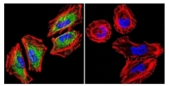
- Experimental details
- Immunofluorescent analysis of Glucocorticoid Receptor using Glucocorticoid Receptor Polyclonal Antibody Cat.-No SP5211P shows staining in 293Cells. Glucocorticoid Receptor (green), F-Actin staining with Phalloidin (red) and nuclei with DAPI (blue) is shown. Cells were grown on chamber slides and fixed with formaldehyde prior to staining. Cells were probed without (control) or with an antibody recognizing Glucocorticoid Receptor Cat.-No SP5211P at a dilution of 1/100 over night at 4°C, washed with PBS and incubated with a DyLight-488 conjugated secondary antibody. Images wee taken at 60x magnification.
Supportive validation
- Submitted by
- Acris Antibodies GmbH (provider)
- Main image

- Experimental details
- Immunohistochemical analysis of Thamnophis sirtalis parietalis (red-sided garter snake) brains using Glucocorticoid Receptor antibody Cat.-No SP5211P.
