Antibody data
- Antibody Data
- Antigen structure
- References [25]
- Comments [0]
- Validations
- Western blot [1]
- Immunocytochemistry [5]
- Immunohistochemistry [1]
- Other assay [5]
Submit
Validation data
Reference
Comment
Report error
- Product number
- PA1-510A - Provider product page

- Provider
- Invitrogen Antibodies
- Product name
- Glucocorticoid Receptor Polyclonal Antibody
- Antibody type
- Polyclonal
- Antigen
- Synthetic peptide
- Description
- PA1-510A detects glucocorticoid receptor (GR) from human, rat, and reptile tissues. This product detects both the unactivated and activated forms of the receptor. PA1-510A has been successfully used in Western blot, immunohistochemistry, immunocytochemistry, immunoprecipitation, immunofluorescence, and gel shift procedures. By Western blot, this antibody detects an ~97 kDa protein representing GR from rat liver extract. PA1-510A immunizing peptide corresponds to amino acid residues 150- 175 from human GR.
- Reactivity
- Human, Mouse, Rat
- Host
- Rabbit
- Isotype
- IgG
- Vial size
- 200 µL
- Concentration
- Conc. Not Determined
- Storage
- -20° C, Avoid Freeze/Thaw Cycles
Submitted references Genome-Wide Identification of Basic Helix-Loop-Helix and NF-1 Motifs Underlying GR Binding Sites in Male Rat Hippocampus.
Reprogramming the chromatin landscape: interplay of the estrogen and glucocorticoid receptors at the genomic level.
Enhanced stimulus sequence-dependent repeated learning in male offspring after prenatal stress alone or in conjunction with lead exposure.
Decreased levels of nuclear glucocorticoid receptor protein in the hippocampus of aged Long-Evans rats with cognitive impairment.
Transcription factor AP1 potentiates chromatin accessibility and glucocorticoid receptor binding.
Distribution and subcellular localization of glucocorticoid receptor-immunoreactive neurons in the developing and adult male zebra finch brain.
Intratympanic dexamethasone as initial therapy for idiopathic sudden sensorineural hearing loss: Clinical evaluation and laboratory investigation.
Administration of glucocorticoids to ovarian cancer patients is associated with expression of the anti-apoptotic genes SGK1 and MKP1/DUSP1 in ovarian tissues.
Characterization of the angiotensin (AT1b) receptor promoter and its regulation by glucocorticoids.
Changes of glucocorticoid receptor expression in the nasal polyps of patients with chronic sinusitis following treatment with glucocorticoid.
Opposite effects of CBP and p300 in glucocorticoid signaling in astrocytes.
Anthrax lethal toxin represses glucocorticoid receptor (GR) transactivation by inhibiting GR-DNA binding in vivo.
Interactions of chronic lead exposure and intermittent stress: consequences for brain catecholamine systems and associated behaviors and HPA axis function.
Involvement of {beta}-catenin and unusual behavior of CBP and p300 in glucocorticosteroid signaling in Schwann cells.
Effects of glucocorticoids on the growth and chemosensitivity of carcinoma cells are heterogeneous and require high concentration of functional glucocorticoid receptors.
NRIP, a novel nuclear receptor interaction protein, enhances the transcriptional activity of nuclear receptors.
Restructuring the neuronal stress response with anti-glucocorticoid gene delivery.
Selective recruitment of p160 coactivators on glucocorticoid-regulated promoters in Schwann cells.
Intronic hormone response elements mediate regulation of FKBP5 by progestins and glucocorticoids.
The glucocorticoid receptor and the orphan nuclear receptor chicken ovalbumin upstream promoter-transcription factor II interact with and mutually affect each other's transcriptional activities: implications for intermediary metabolism.
CBP recruitment and histone acetylation in differential gene induction by glucocorticoids and progestins.
Transcriptional inhibition of the human insulin receptor gene by aldosterone.
Mutation of isoleucine 747 by a threonine alters the ligand responsiveness of the human glucocorticoid receptor.
Identification of a novel dexamethasone responsive enhancer in the human CYP3A5 gene and its activation in human and rat liver cells.
Identification of the activation-labile gene: a single point mutation in the human glucocorticoid receptor presents as two distinct receptor phenotypes.
Pooley JR, Flynn BP, Grøntved L, Baek S, Guertin MJ, Kershaw YM, Birnie MT, Pellatt A, Rivers CA, Schiltz RL, Hager GL, Lightman SL, Conway-Campbell BL
Endocrinology 2017 May 1;158(5):1486-1501
Endocrinology 2017 May 1;158(5):1486-1501
Reprogramming the chromatin landscape: interplay of the estrogen and glucocorticoid receptors at the genomic level.
Miranda TB, Voss TC, Sung MH, Baek S, John S, Hawkins M, Grøntved L, Schiltz RL, Hager GL
Cancer research 2013 Aug 15;73(16):5130-9
Cancer research 2013 Aug 15;73(16):5130-9
Enhanced stimulus sequence-dependent repeated learning in male offspring after prenatal stress alone or in conjunction with lead exposure.
Cory-Slechta DA, Virgolini MB, Liu S, Weston D
Neurotoxicology 2012 Oct;33(5):1188-202
Neurotoxicology 2012 Oct;33(5):1188-202
Decreased levels of nuclear glucocorticoid receptor protein in the hippocampus of aged Long-Evans rats with cognitive impairment.
Lee SY, Hwang YK, Yun HS, Han JS
Brain research 2012 Oct 10;1478:48-54
Brain research 2012 Oct 10;1478:48-54
Transcription factor AP1 potentiates chromatin accessibility and glucocorticoid receptor binding.
Biddie SC, John S, Sabo PJ, Thurman RE, Johnson TA, Schiltz RL, Miranda TB, Sung MH, Trump S, Lightman SL, Vinson C, Stamatoyannopoulos JA, Hager GL
Molecular cell 2011 Jul 8;43(1):145-55
Molecular cell 2011 Jul 8;43(1):145-55
Distribution and subcellular localization of glucocorticoid receptor-immunoreactive neurons in the developing and adult male zebra finch brain.
Shahbazi M, Schmidt M, Carruth LL
General and comparative endocrinology 2011 Dec 1;174(3):354-61
General and comparative endocrinology 2011 Dec 1;174(3):354-61
Intratympanic dexamethasone as initial therapy for idiopathic sudden sensorineural hearing loss: Clinical evaluation and laboratory investigation.
Fu Y, Zhao H, Zhang T, Chi F
Auris, nasus, larynx 2011 Apr;38(2):165-71
Auris, nasus, larynx 2011 Apr;38(2):165-71
Administration of glucocorticoids to ovarian cancer patients is associated with expression of the anti-apoptotic genes SGK1 and MKP1/DUSP1 in ovarian tissues.
Melhem A, Yamada SD, Fleming GF, Delgado B, Brickley DR, Wu W, Kocherginsky M, Conzen SD
Clinical cancer research : an official journal of the American Association for Cancer Research 2009 May 1;15(9):3196-204
Clinical cancer research : an official journal of the American Association for Cancer Research 2009 May 1;15(9):3196-204
Characterization of the angiotensin (AT1b) receptor promoter and its regulation by glucocorticoids.
Bogdarina IG, King PJ, Clark AJ
Journal of molecular endocrinology 2009 Aug;43(2):73-80
Journal of molecular endocrinology 2009 Aug;43(2):73-80
Changes of glucocorticoid receptor expression in the nasal polyps of patients with chronic sinusitis following treatment with glucocorticoid.
Watanabe S, Suzaki H
In vivo (Athens, Greece) 2008 Jan-Feb;22(1):37-42
In vivo (Athens, Greece) 2008 Jan-Feb;22(1):37-42
Opposite effects of CBP and p300 in glucocorticoid signaling in astrocytes.
Fonte C, Trousson A, Grenier J, Schumacher M, Massaad C
The Journal of steroid biochemistry and molecular biology 2007 May;104(3-5):220-7
The Journal of steroid biochemistry and molecular biology 2007 May;104(3-5):220-7
Anthrax lethal toxin represses glucocorticoid receptor (GR) transactivation by inhibiting GR-DNA binding in vivo.
Webster JI, Sternberg EM
Molecular and cellular endocrinology 2005 Sep 28;241(1-2):21-31
Molecular and cellular endocrinology 2005 Sep 28;241(1-2):21-31
Interactions of chronic lead exposure and intermittent stress: consequences for brain catecholamine systems and associated behaviors and HPA axis function.
Virgolini MB, Chen K, Weston DD, Bauter MR, Cory-Slechta DA
Toxicological sciences : an official journal of the Society of Toxicology 2005 Oct;87(2):469-82
Toxicological sciences : an official journal of the Society of Toxicology 2005 Oct;87(2):469-82
Involvement of {beta}-catenin and unusual behavior of CBP and p300 in glucocorticosteroid signaling in Schwann cells.
Fonte C, Grenier J, Trousson A, Chauchereau A, Lahuna O, Baulieu EE, Schumacher M, Massaad C
Proceedings of the National Academy of Sciences of the United States of America 2005 Oct 4;102(40):14260-5
Proceedings of the National Academy of Sciences of the United States of America 2005 Oct 4;102(40):14260-5
Effects of glucocorticoids on the growth and chemosensitivity of carcinoma cells are heterogeneous and require high concentration of functional glucocorticoid receptors.
Lu YS, Lien HC, Yeh PY, Yeh KH, Kuo ML, Kuo SH, Cheng AL
World journal of gastroenterology 2005 Oct 28;11(40):6373-80
World journal of gastroenterology 2005 Oct 28;11(40):6373-80
NRIP, a novel nuclear receptor interaction protein, enhances the transcriptional activity of nuclear receptors.
Tsai TC, Lee YL, Hsiao WC, Tsao YP, Chen SL
The Journal of biological chemistry 2005 May 20;280(20):20000-9
The Journal of biological chemistry 2005 May 20;280(20):20000-9
Restructuring the neuronal stress response with anti-glucocorticoid gene delivery.
Kaufer D, Ogle WO, Pincus ZS, Clark KL, Nicholas AC, Dinkel KM, Dumas TC, Ferguson D, Lee AL, Winters MA, Sapolsky RM
Nature neuroscience 2004 Sep;7(9):947-53
Nature neuroscience 2004 Sep;7(9):947-53
Selective recruitment of p160 coactivators on glucocorticoid-regulated promoters in Schwann cells.
Grenier J, Trousson A, Chauchereau A, Amazit L, Lamirand A, Leclerc P, Guiochon-Mantel A, Schumacher M, Massaad C
Molecular endocrinology (Baltimore, Md.) 2004 Dec;18(12):2866-79
Molecular endocrinology (Baltimore, Md.) 2004 Dec;18(12):2866-79
Intronic hormone response elements mediate regulation of FKBP5 by progestins and glucocorticoids.
Hubler TR, Scammell JG
Cell stress & chaperones 2004 Autumn;9(3):243-52
Cell stress & chaperones 2004 Autumn;9(3):243-52
The glucocorticoid receptor and the orphan nuclear receptor chicken ovalbumin upstream promoter-transcription factor II interact with and mutually affect each other's transcriptional activities: implications for intermediary metabolism.
De Martino MU, Bhattachryya N, Alesci S, Ichijo T, Chrousos GP, Kino T
Molecular endocrinology (Baltimore, Md.) 2004 Apr;18(4):820-33
Molecular endocrinology (Baltimore, Md.) 2004 Apr;18(4):820-33
CBP recruitment and histone acetylation in differential gene induction by glucocorticoids and progestins.
Lambert JR, Nordeen SK
Molecular endocrinology (Baltimore, Md.) 2003 Jun;17(6):1085-94
Molecular endocrinology (Baltimore, Md.) 2003 Jun;17(6):1085-94
Transcriptional inhibition of the human insulin receptor gene by aldosterone.
Calle C, Campión J, García-Arencibia M, Maestro B, Dávila N
The Journal of steroid biochemistry and molecular biology 2003 Apr;84(5):543-53
The Journal of steroid biochemistry and molecular biology 2003 Apr;84(5):543-53
Mutation of isoleucine 747 by a threonine alters the ligand responsiveness of the human glucocorticoid receptor.
Roux S, Térouanne B, Balaguer P, Jausons-Loffreda N, Pons M, Chambon P, Gronemeyer H, Nicolas JC
Molecular endocrinology (Baltimore, Md.) 1996 Oct;10(10):1214-26
Molecular endocrinology (Baltimore, Md.) 1996 Oct;10(10):1214-26
Identification of a novel dexamethasone responsive enhancer in the human CYP3A5 gene and its activation in human and rat liver cells.
Schuetz JD, Schuetz EG, Thottassery JV, Guzelian PS, Strom S, Sun D
Molecular pharmacology 1996 Jan;49(1):63-72
Molecular pharmacology 1996 Jan;49(1):63-72
Identification of the activation-labile gene: a single point mutation in the human glucocorticoid receptor presents as two distinct receptor phenotypes.
Ashraf J, Thompson EB
Molecular endocrinology (Baltimore, Md.) 1993 May;7(5):631-42
Molecular endocrinology (Baltimore, Md.) 1993 May;7(5):631-42
No comments: Submit comment
Supportive validation
- Submitted by
- Invitrogen Antibodies (provider)
- Main image

- Experimental details
- Western blot analysis was performed on membrane enriched extracts (30 µg lysate) of A549 (Lane 1), MCF7 (Lane 2), T-47D (Lane 3), MDA-MB-231 (Lane 4), HeLa (Lane 5) and tissue extract of Mouse Brain (Lane 6). The blot was probed with Anti-Glucocorticoid Receptor Rabbit Polyclonal Antibody (Product # PA1-510A, 1:1,000 dilution) and detected by chemiluminescence using Goat anti-Rabbit IgG (H+L) Superclonal™ Secondary Antibody, HRP conjugate (Product # A27036, 0.25 µg/mL, 1:4000 dilution). A 94 kDa band corresponding to Glucocorticoid Receptor was observed in the cell lines and tissue tested. Known quantity of protein samples were electrophoresed using Novex® NuPAGE® 4-12 % Bis-Tris gel (Product # NP0321BOX), XCell SureLock™ Electrophoresis System (Product # EI0002) and Novex® Sharp Pre-Stained Protein Standard (Product # LC5800). Resolved proteins were then transferred onto a nitrocellulose membrane with iBlot® 2 Dry Blotting System (Product # IB21001). The membrane was probed with the relevant primary and secondary Antibody using iBind™ Flex Western Starter Kit (Product # SLF2000S). Chemiluminescent detection was performed using Pierce™ ECL Western Blotting Substrate (Product # 32106).
Supportive validation
- Submitted by
- Invitrogen Antibodies (provider)
- Main image

- Experimental details
- Immunolocalization of GR in human lymphocytes using Glucocorticoid Receptor Polyclonal Antibody (Product # PA1-510A).
- Submitted by
- Invitrogen Antibodies (provider)
- Main image

- Experimental details
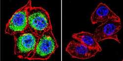
- Immunofluorescent analysis of Glucocorticoid Receptor using Glucocorticoid Receptor Polyclonal Antibody (Product # PA1-510A) shows staining in 293Cells. Glucocorticoid Receptor (green), F-Actin staining with Phalloidin (red) and nuclei with DAPI (blue) is shown. Cells were grown on chamber slides and fixed with formaldehyde prior to staining. Cells were probed without (control) or with an antibody recognizing Glucocorticoid Receptor (Product # PA1-510A) at a dilution of 1:100 over night at 4 °C, washed with PBS and incubated with a DyLight-488 conjugated secondary antibody (Product # 35552 for GAR, Product # 35503 for GAM). Images were taken at 60X magnification.
- Submitted by
- Invitrogen Antibodies (provider)
- Main image

- Experimental details
- Immunofluorescent analysis of Glucocorticoid Receptor using Glucocorticoid Receptor Polyclonal Antibody (Product # PA1-510A) shows staining in A2058 Cells. Glucocorticoid Receptor (green), F-Actin staining with Phalloidin (red) and nuclei with DAPI (blue) is shown. Cells were grown on chamber slides and fixed with formaldehyde prior to staining. Cells were probed without (control) or with an antibody recognizing Glucocorticoid Receptor (Product # PA1-510A) at a dilution of 1:100 over night at 4 °C, washed with PBS and incubated with a DyLight-488 conjugated secondary antibody (Product # 35552 for GAR, Product # 35503 for GAM). Images were taken at 60X magnification.
- Submitted by
- Invitrogen Antibodies (provider)
- Main image

- Experimental details
- Immunofluorescent analysis of Glucocorticoid Receptor using Glucocorticoid Receptor Polyclonal Antibody (Product # PA1-510A) shows staining in Hela Cells. Glucocorticoid Receptor (green), F-Actin staining with Phalloidin (red) and nuclei with DAPI (blue) is shown. Cells were grown on chamber slides and fixed with formaldehyde prior to staining. Cells were probed without (control) or with an antibody recognizing Glucocorticoid Receptor (Product # PA1-510A) at a dilution of 1:100 over night at 4 °C, washed with PBS and incubated with a DyLight-488 conjugated secondary antibody (Product # 35552 for GAR, Product # 35503 for GAM). Images were taken at 60X magnification.
- Submitted by
- Invitrogen Antibodies (provider)
- Main image

- Experimental details
- Immunofluorescence analysis of Glucocorticoid Receptor was performed using 70% confluent log phase A-549 cells. The cells were fixed with 4% paraformaldehyde for 10 minutes, permeabilized with 0.1% Triton™ X-100 for 10 minutes, and blocked with 1% BSA for 1 hour at room temperature. The cells were labeled with Glucocorticoid Receptor Rabbit Polyclonal Antibody (Product # PA1-510A) at 1:250 dilution in 0.1% BSA and incubated for 3 hours at room temperature and then labeled with Goat anti-Rabbit IgG (H+L) Superclonal™ Secondary Antibody, Alexa Fluor® 488 conjugate (Product # A27034) at a dilution of 1:2000 for 45 minutes at room temperature (Panel a: green). Nuclei (Panel b: blue) were stained with SlowFade® Gold Antifade Mountant with DAPI (Product # S36938). F-actin (Panel c: red) was stained with Rhodamine Phalloidin (Product # R415, 1:300). Panel d represents the merged image showing nuclear and cytoplasmic localization. Panel e shows the no primary antibody control. The images were captured at 60X magnification.
Supportive validation
- Submitted by
- Invitrogen Antibodies (provider)
- Main image

- Experimental details
- Immunohistochemical analysis of Thamnophis sirtalis parietalis (red-sided garter snake) brains using Glucocorticoid Receptor Polyclonal Antibody (Product # PA1-510A). Brain tissues were blocked in 10% goat serum in 0.1M phosphate buffered saline, and then stained with PA1-510A at 1:250 dilution for 48 hours at 4°C followed by a biotin-conjugated goat anti-rabbit IgG secondary antibody for 60 minutes at room temperature and then an avidin-biotin complex reagent (60min). Staining was developed by incubating tissues in DAB substrate for 10 minutes. Data courtesy of the Innovators Program.
Supportive validation
- Submitted by
- Invitrogen Antibodies (provider)
- Main image

- Experimental details
- NULL
- Submitted by
- Invitrogen Antibodies (provider)
- Main image

- Experimental details
- NULL
- Submitted by
- Invitrogen Antibodies (provider)
- Main image

- Experimental details
- NULL
- Submitted by
- Invitrogen Antibodies (provider)
- Main image

- Experimental details
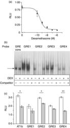
- Figure 4 Glucocorticoid responsiveness of the proximal promoter. (a) The full length promoter-luciferase reporter shows a dose dependent inhibition by dexamethasone in Y1 cells. (b) EMSA with a consensus GRE, or GRE1-4 from the Agtr1b shows a similar complex formation (arrowed) which is not significantly altered by the pre-treatment of cells with dexamethasone. All of the Agtr1b GRE probes show successful competition by a consensus GRE. (c) The Agtr1b promoter reporter still shows adequate function in the presence of mutations of each of the GREs, and glucocorticoid inhibition is retained with the GRE2 and GRE4 mutants, but is lost when GRE1 and 3 are mutated. RLU, relative light units. * P
- Submitted by
- Invitrogen Antibodies (provider)
- Main image

- Experimental details
- Figure 5 Characterization of AP1 sites. (a) EMSA demonstrating that complexes formed on a consensus AP1 sequence and AP1-2 migrate as a complex with similar mobility, and that a consensus oligonucleotide effectively competes with AP1-2 for binding. Note that the mutant AP1 sequence also reveals a non-specific doublet complex of greater mobility than that formed with AP1. (b) EMSA demonstrating that complexes formed on AP1-1 and a consensus AP1 oligonucleotide migrate with similar mobility, but that AP1-1 binding is competed by a consensus oligonucleotide or by the GRE4 oligonucleotide, but not by a mutant consensus AP1 fragment or the other GRE oligonucleotides. (c) The GRE4 probe binds a complex that is supershifted by glucocorticoid receptor antibody (arrow labeled ss), similar to that observed with the consensus GR probe. (d) The AP1-1 probe binds a complex that is supershifted with anti- c-jun and anti-GR antibodies, and to a lesser extent with anti- c-fos antibody.
 Explore
 Validate
 Learn
Learn Western blot
 Immunoprecipitation