Explore
 Validate
 Learn
Learn Western blot
Antibody data
- Antibody Data
- Antigen structure
- References [1]
- Comments [0]
- Validations
- Western blot [2]
- Immunocytochemistry [1]
Submit
Validation data
Reference
Comment
Report error
- Product number
- AF3589 - Provider product page

- Provider
- R&D Systems
- Product name
- Human/Mouse/Rat Cyclophilin A Antibody
- Antibody type
- Polyclonal
- Description
- Antigen Affinity-purified. Detects human, mouse, and rat Cyclophilin A in Western blots.
- Reactivity
- Human, Mouse, Rat
- Host
- Goat
- Conjugate
- Unconjugated
- Antigen sequence
P62937- Isotype
- IgG
- Vial size
- 100 ug
- Concentration
- LYOPH
- Storage
- Use a manual defrost freezer and avoid repeated freeze-thaw cycles. 12 months from date of receipt, -20 to -70 °C as supplied. 1 month, 2 to 8 °C under sterile conditions after reconstitution. 6 months, -20 to -70 °C under sterile conditions after reconstitution.
Submitted references The Novel Extracellular Cyclophilin A (CyPA) - Inhibitor MM284 Reduces Myocardial Inflammation and Remodeling in a Mouse Model of Troponin I -Induced Myocarditis.
Heinzmann D, Bangert A, Müller AM, von Ungern-Sternberg SN, Emschermann F, Schönberger T, Chatterjee M, Mack AF, Klingel K, Kandolf R, Malesevic M, Borst O, Gawaz M, Langer HF, Katus H, Fischer G, May AE, Kaya Z, Seizer P
PloS one 2015;10(4):e0124606
PloS one 2015;10(4):e0124606
No comments: Submit comment
Supportive validation
- Submitted by
- R&D Systems (provider)
- Main image
- Experimental details
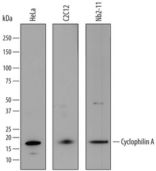
- Detection of Human, Mouse, and Rat Cyclophilin A by Western Blot. Western blot shows lysates of HeLa human cervical epithelial carcinoma cell line, C2C12 mouse myoblast cell line, and Nb2-11 rat lymphoma cell line. PVDF membrane was probed with 0.1 µg/mL of Goat Anti-Human Cyclophilin A Antigen Affinity-purified Polyclonal Antibody (Catalog # AF3589) followed by HRP-conjugated Anti-Goat IgG Secondary Antibody (Catalog # HAF017). A specific band was detected for Cyclophilin A at approximately 18 kDa (as indicated). This experiment was conducted under reducing conditions and using Immunoblot Buffer Group 1.

- Submitted by
- R&D Systems (provider)
- Main image

- Experimental details
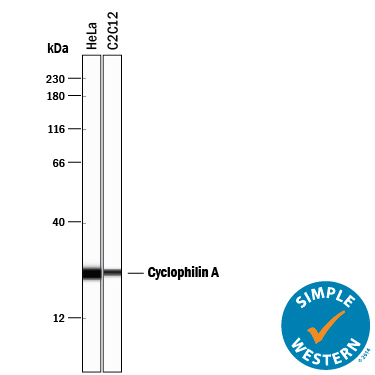
- Detection of Human and Mouse Cyclophilin A by Simple WesternTM. Simple Western lane view shows lysates of HeLa human cervical epithelial carcinoma cell line and C2C12 mouse myoblast cell line, loaded at 0.2 mg/mL. A specific band was detected for Cyclophilin A at approximately 25 kDa (as indicated) using 5 µg/mL of Goat Anti-Human/Mouse/Rat Cyclophilin A Antigen Affinity-purified Polyclonal Antibody (Catalog # AF3589) followed by 1:50 dilution of HRP-conjugated Anti-Goat IgG Secondary Antibody (Catalog # HAF109). This experiment was conducted under reducing conditions and using the 12-230 kDa separation system.
Supportive validation
- Submitted by
- R&D Systems (provider)
- Main image

- Experimental details
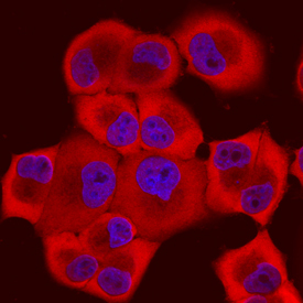
- Cyclophilin A in PANC-1 Human Cell Line. Cyclophilin A was detected in immersion fixed PANC-1 human pancreatic carcinoma cell line using Goat Anti-Human/Mouse/Rat Cyclophilin A Antigen Affinity-purified Polyclonal Antibody (Catalog # AF3589) at 10 µg/mL for 3 hours at room temperature. Cells were stained using the Northern-Lights™ 557-conjugated Anti-Goat IgG Secondary Antibody (red; Catalog # NL001) and counterstained with DAPI (blue). Specific staining was localized to nuclei and cytoplasm. View our protocol for Fluorescent ICC Staining of Cells on Coverslips.