Explore
 Validate
 Learn
Learn Western blot
 Flow cytometry
Antibody data
- Antibody Data
- Antigen structure
- References [1]
- Comments [0]
- Validations
- Western blot [3]
- Immunocytochemistry [1]
- Immunoprecipitation [1]
- Immunohistochemistry [2]
Submit
Validation data
Reference
Comment
Report error
- Product number
- GTX629691 - Provider product page

- Provider
- GeneTex
- Product name
- E-Cadherin antibody [GT477]
- Antibody type
- Monoclonal
- Reactivity
- Human, Mouse, Rat
- Host
- Mouse
Submitted references Methyl-CpG Binding Domain Protein 2 Inhibits the Malignant Characteristic of Lung Adenocarcinoma through the Epigenetic Modulation of 10 to 11 Translocation 1 and miR-200s.
Pei YF, Xu XN, Wang ZF, Wang FW, Wu WD, Geng JF, Liu XQ
The American journal of pathology 2019 Feb 5;
The American journal of pathology 2019 Feb 5;
No comments: Submit comment
Supportive validation
- Submitted by
- GeneTex (provider)
- Main image

- Experimental details
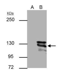
- E-cadherin antibody [GT477] detects E-cadherin protein by Western blot analysis.A. 30 £gg MCF-7 whole cell lysate/extractB. 30 £gg MDA-MB-231 whole cell lysate/extract5 % SDS-PAGEE-cadherin antibody [GT477] (GTX629691) dilution: 1:1000

- Submitted by
- GeneTex (provider)
- Main image
- Experimental details
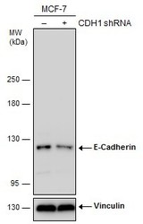
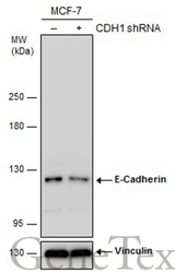
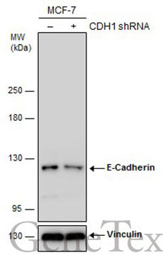
- Non-transfected (¡V) and transfected (+) MCF-7 whole cell extracts (30 ?g) were separated by 5% SDS-PAGE, and the membrane was blotted with E-Cadherin antibody [GT477] (GTX629691) diluted at 1:2000. The HRP-conjugated anti-mouset IgG antibody (GTX213111-01) was used to detect the primary antibody.
- Submitted by
- GeneTex (provider)
- Main image
- Experimental details
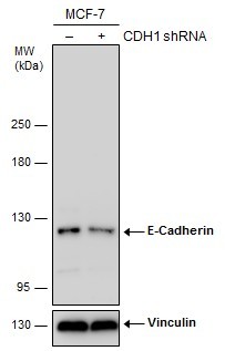
- Non-transfected (¡V) and transfected (+) MCF-7 whole cell extracts (30 ?g) were separated by 5% SDS-PAGE, and the membrane was blotted with E-Cadherin antibody [GT477] (GTX629691) diluted at 1:2000. The HRP-conjugated anti-mouset IgG antibody (GTX213111-01) was used to detect the primary antibody.
Supportive validation
- Submitted by
- GeneTex (provider)
- Main image
- Experimental details
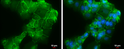
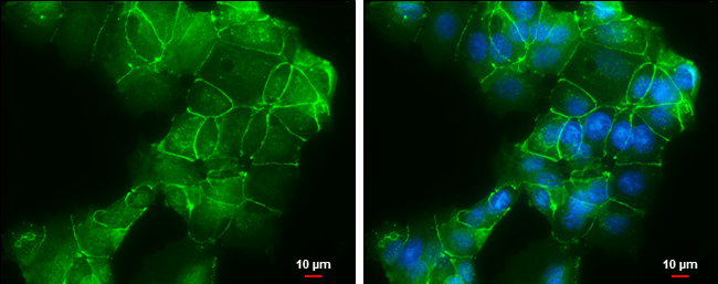
- E-Cadherin antibody [GT477] detects E-Cadherin protein at cell membrane by immunofluorescent analysis.Sample: MCF7 cells were fixed in ice-cold MeOH for 5 min.Green: E-Cadherin protein stained by E-Cadherin antibody [GT477] (GTX629691) diluted at 1:500.Blue: Hoechst 33342 staining.Scale bar = 10 £gm.
Supportive validation
- Submitted by
- GeneTex (provider)
- Main image
- Experimental details
- E-adherin antibody [GT477] immunoprecipitates E-adherin protein in IP experiments.IP samples: MCF-7 whole cell extractA. Control with 3 £gg of preimmune Mouse IgGB. Immunoprecipitation of E-adherin protein by 3 £gg E-adherin antibody [GT477] (GTX629691)5 % SDS-PAGEThe immunoprecipitated E-adherin protein was detected by E-adherin antibody [GT477] (GTX629691) diluted at 1:500.[EasyBlot anti-mouse IgG (GTX221667-01) was used as a secondary reagent]

Supportive validation
- Submitted by
- GeneTex (provider)
- Main image
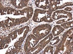
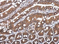
- Experimental details
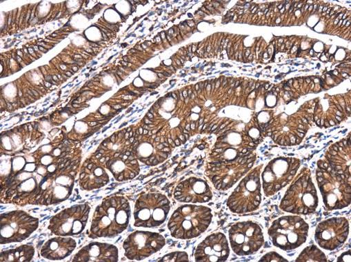
- E-Cadherin antibody [GT477] detects E-Cadherin protein at cell membrane and cytoplasm in mouse intestine by immunohistochemical analysis. Sample: Paraffin-embedded mouse intestine. E-Cadherin antibody [GT477] (GTX629691) diluted at 1:250.
- Submitted by
- GeneTex (provider)
- Main image
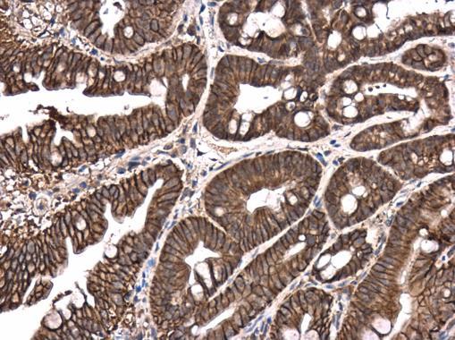
- Experimental details
- E-Cadherin antibody [GT477] detects E-Cadherin protein at cell membrane and cytoplasm in rat intestine by immunohistochemical analysis. Sample: Paraffin-embedded rat intestine. E-Cadherin antibody [GT477] (GTX629691) diluted at 1:250.