Explore
 Validate
 Learn
Learn Western blot
 Immunocytochemistry
Antibody data
- Antibody Data
- Antigen structure
- References [31]
- Comments [0]
- Validations
- Western blot [10]
- Immunocytochemistry [3]
- Immunoprecipitation [2]
- Immunohistochemistry [8]
Submit
Validation data
Reference
Comment
Report error
- Product number
- GTX100443 - Provider product page

- Provider
- GeneTex
- Proper citation
- GeneTex Cat#GTX100443, RRID:AB_10729586
- Product name
- E-Cadherin antibody
- Antibody type
- Polyclonal
- Reactivity
- Human, Mouse, Rat, Zebrafish
- Host
- Rabbit
Submitted references Sympathetic innervation contributes to perineural invasion of salivary adenoid cystic carcinoma via the β2-adrenergic receptor.
ZEB2, a master regulator of the epithelial-mesenchymal transition, mediates trophoblast differentiation.
Prickle1 is required for EMT and migration of zebrafish cranial neural crest.
PEGylated liposome-encapsulated rhenium-188 radiopharmaceutical inhibits proliferation and epithelial-mesenchymal transition of human head and neck cancer cells in vivo with repeated therapy.
Upregulation of LGALS1 is associated with oral cancer metastasis.
Profiling human breast epithelial cells using single cell RNA sequencing identifies cell diversity.
Activation of fibroblasts by nicotine promotes the epithelial-mesenchymal transition and motility of breast cancer cells.
The kinesin Eg5 inhibitor K858 induces apoptosis and reverses the malignant invasive phenotype in human glioblastoma cells.
Human trophoblast epithelial-mesenchymal transition in abnormally invasive placenta.
Glycine Is a Predictor for a Suppressive Effect of Fucoxanthinol on Colonosphere Formation Under Hypoxia.
Effects of bradykinin on TGF‑β1‑induced epithelial‑mesenchymal transition in ARPE‑19 cells.
Low-Dose Alkylphenol Exposure Promotes Mammary Epithelium Alterations and Transgenerational Developmental Defects, But Does Not Enhance Tumorigenic Behavior of Breast Cancer Cells.
Hypoxia and TGF-β1 induced PLOD2 expression improve the migration and invasion of cervical cancer cells by promoting epithelial-to-mesenchymal transition (EMT) and focal adhesion formation.
Reverse epithelial-mesenchymal transition contributes to the regain of drug sensitivity in tyrosine kinase inhibitor-resistant non-small cell lung cancer cells.
Mammary epithelial cell phenotype disruption in vitro and in vivo through ERalpha36 overexpression.
Knockdown of IQGAP1 inhibits proliferation and epithelial-mesenchymal transition by Wnt/β-catenin pathway in thyroid cancer.
Runx3 is a key modulator during the epithelial-mesenchymal transition of alveolar type II cells in animal models of BPD.
Cytoplasmic aryl hydrocarbon receptor regulates glycogen synthase kinase 3 beta, accelerates vimentin degradation, and suppresses epithelial-mesenchymal transition in non-small cell lung cancer cells.
Sulfiredoxin may promote metastasis and invasion of cervical squamous cell carcinoma by epithelial-mesenchymal transition.
Downregulation of miR-199a/b-5p is associated with GCNT2 induction upon epithelial-mesenchymal transition in colon cancer.
The inhibition of lung cancer cell migration by AhR-regulated autophagy.
Transcription factor SPZ1 promotes TWIST-mediated epithelial-mesenchymal transition and oncogenesis in human liver cancer.
Metronomic Cordycepin Therapy Prolongs Survival of Oral Cancer-Bearing Mice and Inhibits Epithelial-Mesenchymal Transition.
Oxystressed tumor microenvironment potentiates epithelial to mesenchymal transition and alters cellular bioenergetics towards cancer progression.
miR-204 inhibits invasion and epithelial-mesenchymal transition by targeting FOXM1 in esophageal cancer.
Targeting annexin A2 reduces tumorigenesis and therapeutic resistance of nasopharyngeal carcinoma.
Long non-coding RNA AOC4P suppresses hepatocellular carcinoma metastasis by enhancing vimentin degradation and inhibiting epithelial-mesenchymal transition.
PGRMC1 contributes to doxorubicin-induced chemoresistance in MES-SA uterine sarcoma.
Comprehensive proteome quantification reveals NgBR as a new regulator for epithelial-mesenchymal transition of breast tumor cells.
Micro-scaffold array chip for upgrading cell-based high-throughput drug testing to 3D using benchtop equipment.
CD63 tetraspanin is a negative driver of epithelial-to-mesenchymal transition in human melanoma cells.
Ma C, Gao T, Ju J, Zhang Y, Ni Q, Li Y, Zhao Z, Chai J, Yang X, Sun M
OncoTargets and therapy 2019;12:1475-1495
OncoTargets and therapy 2019;12:1475-1495
ZEB2, a master regulator of the epithelial-mesenchymal transition, mediates trophoblast differentiation.
DaSilva-Arnold SC, Kuo CY, Davra V, Remache Y, Kim PCW, Fisher JP, Zamudio S, Al-Khan A, Birge RB, Illsley NP
Molecular human reproduction 2019 Feb 1;25(2):61-75
Molecular human reproduction 2019 Feb 1;25(2):61-75
Prickle1 is required for EMT and migration of zebrafish cranial neural crest.
Ahsan K, Singh N, Rocha M, Huang C, Prince VE
Developmental biology 2019 Apr 1;448(1):16-35
Developmental biology 2019 Apr 1;448(1):16-35
PEGylated liposome-encapsulated rhenium-188 radiopharmaceutical inhibits proliferation and epithelial-mesenchymal transition of human head and neck cancer cells in vivo with repeated therapy.
Chang CY, Chen CC, Lin LT, Chang CH, Chen LC, Wang HE, Lee TW, Lee YJ
Cell death discovery 2018;4:100
Cell death discovery 2018;4:100
Upregulation of LGALS1 is associated with oral cancer metastasis.
Li JM, Tseng CW, Lin CC, Law CH, Chien YA, Kuo WH, Chou HC, Wang WC, Chan HL
Therapeutic advances in medical oncology 2018;10:1758835918794622
Therapeutic advances in medical oncology 2018;10:1758835918794622
Profiling human breast epithelial cells using single cell RNA sequencing identifies cell diversity.
Nguyen QH, Pervolarakis N, Blake K, Ma D, Davis RT, James N, Phung AT, Willey E, Kumar R, Jabart E, Driver I, Rock J, Goga A, Khan SA, Lawson DA, Werb Z, Kessenbrock K
Nature communications 2018 May 23;9(1):2028
Nature communications 2018 May 23;9(1):2028
Activation of fibroblasts by nicotine promotes the epithelial-mesenchymal transition and motility of breast cancer cells.
Chen PC, Lee WY, Ling HH, Cheng CH, Chen KC, Lin CW
Journal of cellular physiology 2018 Jun;233(6):4972-4980
Journal of cellular physiology 2018 Jun;233(6):4972-4980
The kinesin Eg5 inhibitor K858 induces apoptosis and reverses the malignant invasive phenotype in human glioblastoma cells.
Taglieri L, Rubinacci G, Giuffrida A, Carradori S, Scarpa S
Investigational new drugs 2018 Feb;36(1):28-35
Investigational new drugs 2018 Feb;36(1):28-35
Human trophoblast epithelial-mesenchymal transition in abnormally invasive placenta.
DaSilva-Arnold SC, Zamudio S, Al-Khan A, Alvarez-Perez J, Mannion C, Koenig C, Luke D, Perez AM, Petroff M, Alvarez M, Illsley NP
Biology of reproduction 2018 Aug 1;99(2):409-421
Biology of reproduction 2018 Aug 1;99(2):409-421
Glycine Is a Predictor for a Suppressive Effect of Fucoxanthinol on Colonosphere Formation Under Hypoxia.
Terasaki M, Ogawa Y, Endo T, Maeda H, Hamada J, Osada K, Miyashita K, Mutoh M
Anticancer research 2018 Apr;38(4):2169-2179
Anticancer research 2018 Apr;38(4):2169-2179
Effects of bradykinin on TGF‑β1‑induced epithelial‑mesenchymal transition in ARPE‑19 cells.
Wei Q, Liu Q, Ren C, Liu J, Cai W, Zhu M, Jin H, He M, Yu J
Molecular medicine reports 2018 Apr;17(4):5878-5886
Molecular medicine reports 2018 Apr;17(4):5878-5886
Low-Dose Alkylphenol Exposure Promotes Mammary Epithelium Alterations and Transgenerational Developmental Defects, But Does Not Enhance Tumorigenic Behavior of Breast Cancer Cells.
Chamard-Jovenin C, Thiebaut C, Chesnel A, Bresso E, Morel C, Smail-Tabbone M, Devignes MD, Boukhobza T, Dumond H
Frontiers in endocrinology 2017;8:272
Frontiers in endocrinology 2017;8:272
Hypoxia and TGF-β1 induced PLOD2 expression improve the migration and invasion of cervical cancer cells by promoting epithelial-to-mesenchymal transition (EMT) and focal adhesion formation.
Xu F, Zhang J, Hu G, Liu L, Liang W
Cancer cell international 2017;17:54
Cancer cell international 2017;17:54
Reverse epithelial-mesenchymal transition contributes to the regain of drug sensitivity in tyrosine kinase inhibitor-resistant non-small cell lung cancer cells.
Lee AF, Chen MC, Chen CJ, Yang CJ, Huang MS, Liu YP
PloS one 2017;12(7):e0180383
PloS one 2017;12(7):e0180383
Mammary epithelial cell phenotype disruption in vitro and in vivo through ERalpha36 overexpression.
Thiebaut C, Chamard-Jovenin C, Chesnel A, Morel C, Djermoune EH, Boukhobza T, Dumond H
PloS one 2017;12(3):e0173931
PloS one 2017;12(3):e0173931
Knockdown of IQGAP1 inhibits proliferation and epithelial-mesenchymal transition by Wnt/β-catenin pathway in thyroid cancer.
Su D, Liu Y, Song T
OncoTargets and therapy 2017;10:1549-1559
OncoTargets and therapy 2017;10:1549-1559
Runx3 is a key modulator during the epithelial-mesenchymal transition of alveolar type II cells in animal models of BPD.
Yang H, Fu J, Yao L, Hou A, Xue X
International journal of molecular medicine 2017 Nov;40(5):1466-1476
International journal of molecular medicine 2017 Nov;40(5):1466-1476
Cytoplasmic aryl hydrocarbon receptor regulates glycogen synthase kinase 3 beta, accelerates vimentin degradation, and suppresses epithelial-mesenchymal transition in non-small cell lung cancer cells.
Li CH, Liu CW, Tsai CH, Peng YJ, Yang YH, Liao PL, Lee CC, Cheng YW, Kang JJ
Archives of toxicology 2017 May;91(5):2165-2178
Archives of toxicology 2017 May;91(5):2165-2178
Sulfiredoxin may promote metastasis and invasion of cervical squamous cell carcinoma by epithelial-mesenchymal transition.
Chen X, Lan K, Liu Q, Yang X, Wang H
Tumour biology : the journal of the International Society for Oncodevelopmental Biology and Medicine 2017 Mar;39(3):1010428317695942
Tumour biology : the journal of the International Society for Oncodevelopmental Biology and Medicine 2017 Mar;39(3):1010428317695942
Downregulation of miR-199a/b-5p is associated with GCNT2 induction upon epithelial-mesenchymal transition in colon cancer.
Chao CC, Wu PH, Huang HC, Chung HY, Chou YC, Cai BH, Kannagi R
FEBS letters 2017 Jul;591(13):1902-1917
FEBS letters 2017 Jul;591(13):1902-1917
The inhibition of lung cancer cell migration by AhR-regulated autophagy.
Tsai CH, Li CH, Cheng YW, Lee CC, Liao PL, Lin CH, Huang SH, Kang JJ
Scientific reports 2017 Feb 14;7:41927
Scientific reports 2017 Feb 14;7:41927
Transcription factor SPZ1 promotes TWIST-mediated epithelial-mesenchymal transition and oncogenesis in human liver cancer.
Wang LT, Chiou SS, Chai CY, Hsi E, Chiang CM, Huang SK, Wang SN, Yokoyama KK, Hsu SH
Oncogene 2017 Aug;36(31):4405-4414
Oncogene 2017 Aug;36(31):4405-4414
Metronomic Cordycepin Therapy Prolongs Survival of Oral Cancer-Bearing Mice and Inhibits Epithelial-Mesenchymal Transition.
Su NW, Wu SH, Chi CW, Liu CJ, Tsai TH, Chen YJ
Molecules (Basel, Switzerland) 2017 Apr 13;22(4)
Molecules (Basel, Switzerland) 2017 Apr 13;22(4)
Oxystressed tumor microenvironment potentiates epithelial to mesenchymal transition and alters cellular bioenergetics towards cancer progression.
Sridaran D, Ramamoorthi G, MahaboobKhan R, Kumpati P
Tumour biology : the journal of the International Society for Oncodevelopmental Biology and Medicine 2016 Oct;37(10):13307-13322
Tumour biology : the journal of the International Society for Oncodevelopmental Biology and Medicine 2016 Oct;37(10):13307-13322
miR-204 inhibits invasion and epithelial-mesenchymal transition by targeting FOXM1 in esophageal cancer.
Sun Y, Yu X, Bai Q
International journal of clinical and experimental pathology 2015;8(10):12775-83
International journal of clinical and experimental pathology 2015;8(10):12775-83
Targeting annexin A2 reduces tumorigenesis and therapeutic resistance of nasopharyngeal carcinoma.
Chen CY, Lin YS, Chen CL, Chao PZ, Chiou JF, Kuo CC, Lee FP, Lin YF, Sung YH, Lin YT, Li CF, Chen YJ, Chen CH
Oncotarget 2015 Sep 29;6(29):26946-59
Oncotarget 2015 Sep 29;6(29):26946-59
Long non-coding RNA AOC4P suppresses hepatocellular carcinoma metastasis by enhancing vimentin degradation and inhibiting epithelial-mesenchymal transition.
Wang TH, Lin YS, Chen Y, Yeh CT, Huang YL, Hsieh TH, Shieh TM, Hsueh C, Chen TC
Oncotarget 2015 Sep 15;6(27):23342-57
Oncotarget 2015 Sep 15;6(27):23342-57
PGRMC1 contributes to doxorubicin-induced chemoresistance in MES-SA uterine sarcoma.
Lin ST, May EW, Chang JF, Hu RY, Wang LH, Chan HL
Cellular and molecular life sciences : CMLS 2015 Jun;72(12):2395-409
Cellular and molecular life sciences : CMLS 2015 Jun;72(12):2395-409
Comprehensive proteome quantification reveals NgBR as a new regulator for epithelial-mesenchymal transition of breast tumor cells.
Zhao B, Xu B, Hu W, Song C, Wang F, Liu Z, Ye M, Zou H, Miao QR
Journal of proteomics 2015 Jan 1;112:38-52
Journal of proteomics 2015 Jan 1;112:38-52
Micro-scaffold array chip for upgrading cell-based high-throughput drug testing to 3D using benchtop equipment.
Li X, Zhang X, Zhao S, Wang J, Liu G, Du Y
Lab on a chip 2014 Feb 7;14(3):471-81
Lab on a chip 2014 Feb 7;14(3):471-81
CD63 tetraspanin is a negative driver of epithelial-to-mesenchymal transition in human melanoma cells.
Lupia A, Peppicelli S, Witort E, Bianchini F, Carloni V, Pimpinelli N, Urso C, Borgognoni L, Capaccioli S, Calorini L, Lulli M
The Journal of investigative dermatology 2014 Dec;134(12):2947-2956
The Journal of investigative dermatology 2014 Dec;134(12):2947-2956
No comments: Submit comment
Enhanced validation
Supportive validation
- Submitted by
- GeneTex (provider)
- Enhanced method
- Genetic validation
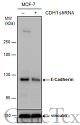
- Main image
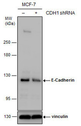
- Experimental details
- Non-transfected (¡V) and transfected (+) MCF-7 whole cell extracts (30 ?g) were separated by 5% SDS-PAGE, and the membrane was blotted with E-Cadherin antibody (GTX100443) diluted at 1:7000. The HRP-conjugated anti-rabbit IgG antibody (GTX213110-01) was used to detect the primary antibody.
Supportive validation
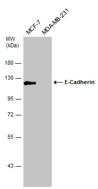
- Submitted by
- GeneTex (provider)
- Main image

- Experimental details
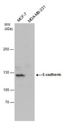
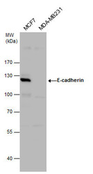
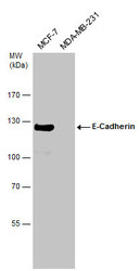
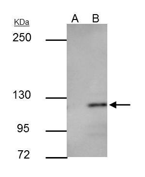
- E-cadherin antibody detects E-cadherin protein by Western blot analysis.A. 30 ?g MCF-7 whole cell lysate/extractB. 30 ?g MDA-MB-231 whole cell lysate/extract5 % SDS-PAGEE-cadherin antibody (GTX100443) dilution: 1:2000
- Validation comment
- WB
- Submitted by
- GeneTex (provider)
- Main image
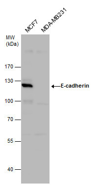
- Experimental details
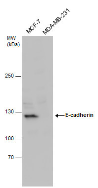
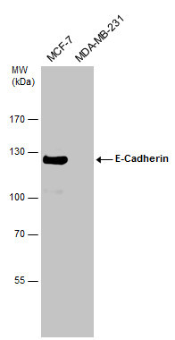
- E-cadherin antibody detects E-cadherin protein by western blot analysis.A. 30 ?g MCF-7 whole cell lysate/extractB. 30 ?g MDA-MB-231 whole cell lysate/extract5 % SDS-PAGEE-cadherin antibody (GTX100443) dilution: 1:2000
- Validation comment
- WB
- Submitted by
- GeneTex (provider)
- Main image
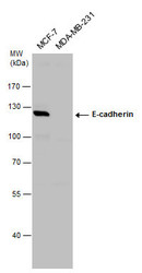
- Experimental details
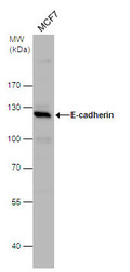
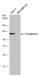
- E-cadherin antibody detects E-cadherin protein by western blot analysis. Various whole cell extracts (30 ?g) were separated by 7.5% SDS-PAGE, and the membrane was blotted with E-cadherin antibody (GTX100443) diluted at a dilution of 1:2000.
- Validation comment
- WB
- Submitted by
- GeneTex (provider)
- Main image
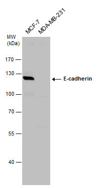
- Experimental details
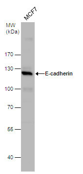
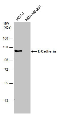
- E-cadherin antibody detects E-cadherin protein by western blot analysis. Various whole cell extracts (30 ?g) were separated by 7.5% SDS-PAGE, and the membrane was blotted with E-cadherin antibody (GTX100443) diluted at a dilution of 1:2000.
- Validation comment
- WB
- Submitted by
- GeneTex (provider)
- Main image
- Experimental details
- E-cadherin antibody detects E-cadherin protein by western blot analysis. Whole cell extracts (30 ?g) was separated by 5% SDS-PAGE, and the membrane was blotted with E-cadherin antibody (GTX100443) at a dilution of 1:2000.
- Validation comment
- WB
- Submitted by
- GeneTex (provider)
- Main image
- Experimental details
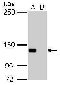
- Various whole cell extracts (30 ?g) were separated by 7.5% SDS-PAGE, and the membrane was blotted with E-Cadherin antibody (GTX100443) diluted at 1:2000. The HRP-conjugated anti-rabbit IgG antibody (GTX213110-01) was used to detect the primary antibody.
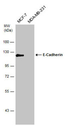
- Submitted by
- GeneTex (provider)
- Main image
- Experimental details
- Various whole cell extracts (30 ?g) were separated by 7.5% SDS-PAGE, and the membrane was blotted with E-Cadherin antibody (GTX100443) diluted at 1:2000.
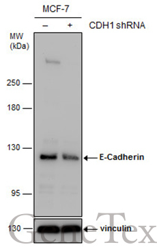
- Submitted by
- GeneTex (provider)
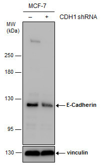
- Main image
- Experimental details
- Non-transfected (¡V) and transfected (+) MCF-7 whole cell extracts (30 ?g) were separated by 5% SDS-PAGE, and the membrane was blotted with E-Cadherin antibody (GTX100443) diluted at 1:7000. The HRP-conjugated anti-rabbit IgG antibody (GTX213110-01) was used to detect the primary antibody.
- Submitted by
- GeneTex (provider)
- Main image
- Experimental details
- Various whole cell extracts (30 ?g) were separated by 7.5% SDS-PAGE, and the membrane was blotted with E-Cadherin antibody (GTX100443) diluted at 1:2000.
Supportive validation
- Submitted by
- GeneTex (provider)
- Main image

- Experimental details
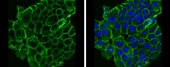
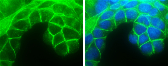
- E-Cadherin antibody detects E-Cadherin protein at cell membrane by immunofluorescent analysis.Sample: A431 cells were fixed in 4% paraformaldehyde at RT for 15 min.Green: E-Cadherin protein stained by E-Cadherin antibody (GTX100443) diluted at 1:500.Blue: Hoechst 33342 staining.
- Submitted by
- GeneTex (provider)
- Main image

- Experimental details
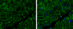
- E-Cadherin antibody detects E-Cadherin protein at cell membrane by immunofluorescent analysis.Sample: MCF7 cells were fixed in 4% paraformaldehyde at RT for 15 min.Green: E-Cadherin protein stained by E-Cadherin antibody (GTX100443) diluted at 1:500.Blue: Hoechst 33342 staining.
- Submitted by
- GeneTex (provider)
- Main image
- Experimental details
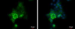
- E-Cadherin antibody detects E-Cadherin protein at cell membrane by immunofluorescent analysis.Sample: HCT 116 cells were fixed in 4% paraformaldehyde at RT for 15 min.Green: E-Cadherin protein stained by E-Cadherin antibody (GTX100443) diluted at 1:500.Blue: Hoechst 33342 staining.Scale bar = 10 £gm.

Supportive validation
- Submitted by
- GeneTex (provider)
- Main image

- Experimental details
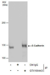
- E-cadherin antibody immunoprecipitates E-cadherin protein in IP experiments.IP samples: MCF-7 whole cell extractA. Control with 3 £gg of preimmune Rabbit IgGB. Immunoprecipitation of E-cadherin protein by 3 £gg E-cadherin antibody (GTX100443)5 % SDS-PAGEThe immunoprecipitated E-cadherin protein was detected by E-cadherin antibody (GTX100443) diluted at 1:500.[EasyBlot anti-rabbit IgG (GTX221666-01) was used as a secondary reagent]
- Submitted by
- GeneTex (provider)
- Main image
- Experimental details
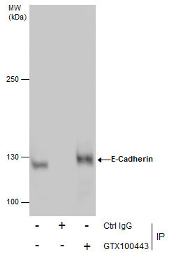
- Immunoprecipitation of E-Cadherin protein from MCF-7 whole cell extracts using 5 £gg of E-Cadherin antibody (GTX100443).Western blot analysis was performed using E-Cadherin antibody (GTX100443).EasyBlot anti-Rabbit IgG (GTX221666-01) was used as a secondary reagent.
Supportive validation
- Submitted by
- GeneTex (provider)
- Main image
- Experimental details
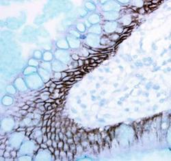
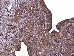
- Immunohistochemical analysis of paraffin-embedded human ulcerative colitis tissue using E-Cadherin antibody (GTX100443).

- Submitted by
- GeneTex (provider)
- Main image
- Experimental details
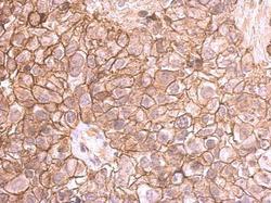
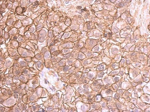
- E-cadherin antibody detects E-cadherin protein at membrane on human breast cancer by immunohistochemical analysis. Sample: Paraffin-embedded breast cancer. E-cadherin antibody (GTX100443) dilution: 1:500.
- Submitted by
- GeneTex (provider)
- Main image
- Experimental details
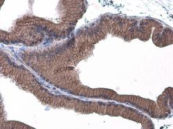
- E-Cadherin antibody detects E-Cadherin protein at cell membrane in mouse pancreas by immunohistochemical analysis. Sample: Paraffin-embedded mouse pancreas. E-Cadherin antibody (GTX100443) diluted at 1:400.

- Submitted by
- GeneTex (provider)
- Main image
- Experimental details
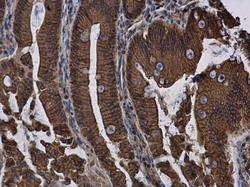
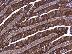
- E-Cadherin antibody detects E-Cadherin protein at cell membrane and cytoplasm in rat duodenum by immunohistochemical analysis. Sample: Paraffin-embedded rat duodenum. E-Cadherin antibody (GTX100443) diluted at 1:500.
- Submitted by
- GeneTex (provider)
- Main image
- Experimental details
- E-Cadherin antibody detects E-Cadherin protein at cell membrane in mouse cervix by immunohistochemical analysis. Sample: Paraffin-embedded mouse cervix. E-Cadherin antibody (GTX100443) diluted at 1:500.

- Submitted by
- GeneTex (provider)
- Main image
- Experimental details
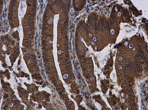
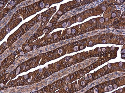
- E-Cadherin antibody detects E-Cadherin protein at cell membrane in rat intestine by immunohistochemical analysis. Sample: Paraffin-embedded rat intestine. E-Cadherin antibody (GTX100443) diluted at 1:500.
- Submitted by
- GeneTex (provider)
- Main image
- Experimental details
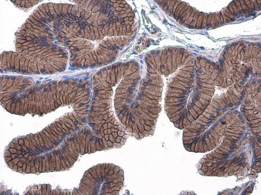
- E-Cadherin antibody detects E-Cadherin protein at cell membrane in rat prostate by immunohistochemical analysis. Sample: Paraffin-embedded rat prostate. E-Cadherin antibody (GTX100443) diluted at 1:500.

- Submitted by
- GeneTex (provider)
- Main image

- Experimental details
- E-Cadherin antibody detects E-Cadherin protein at cell membrane in rat prostate by immunohistochemical analysis. Sample: Paraffin-embedded rat prostate. E-Cadherin antibody (GTX100443) diluted at 1:500.