Explore
 Validate
 Learn
Learn Western blot
 Immunocytochemistry
Antibody data
- Antibody Data
- Antigen structure
- References [1]
- Comments [0]
- Validations
- Western blot [2]
- Immunohistochemistry [16]
Submit
Validation data
Reference
Comment
Report error
- Product number
- UM800076 - Provider product page

- Provider
- OriGene
- Proper citation
- OriGene Cat#UM800076, RRID:AB_2629186
- Product name
- CDH1 mouse monoclonal antibody,clone UMAB184
- Antibody type
- Monoclonal
- Description
- CDH1 mouse monoclonal antibody,clone UMAB184
- Host
- Mouse
- Conjugate
- Unconjugated
- Epitope
- CDH1
- Isotype
- IgG
- Antibody clone number
- UMAB184
- Vial size
- 100 µl
- Concentration
- 1.00mg/ml
Submitted references Prediction model of lymphovascular invasion based on clinicopathological factors in Chinese patients with invasive breast cancer.
Shen S, Wu G, Xiao G, Du R, Hu N, Xia X, Zhou H
Medicine 2018 Oct;97(43):e12973
Medicine 2018 Oct;97(43):e12973
No comments: Submit comment
Supportive validation
- Submitted by
- OriGene (provider)
- Main image
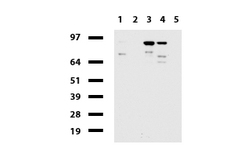
- Experimental details
- Western blot of human tissue lysates (15ug) from 5 different tissues (1: Liver, 2: Ovary, 3: Thyroid, 4: Colon, 5: Spleen ). Diluation: 1:500.
- Validation comment
- WB

- Submitted by
- OriGene (provider)
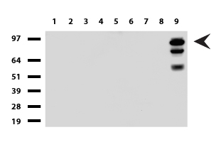
- Main image

- Experimental details
- Western blot of cell lysates (35ug) from 9 different cell lines (1: HepG2, 2: HeLa, 3: SV-T2, 4: A549, 5: COS7, 6: Jurkat, 7: MDCK, 8: PC-12, 9: MCF7). Diluation: 1:500
- Validation comment
- WB
Supportive validation
- Submitted by
- OriGene (provider)
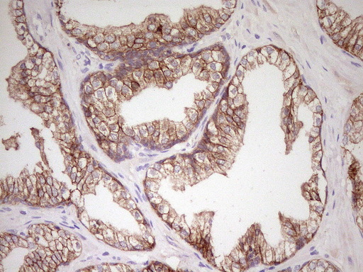
- Main image

- Experimental details
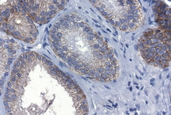
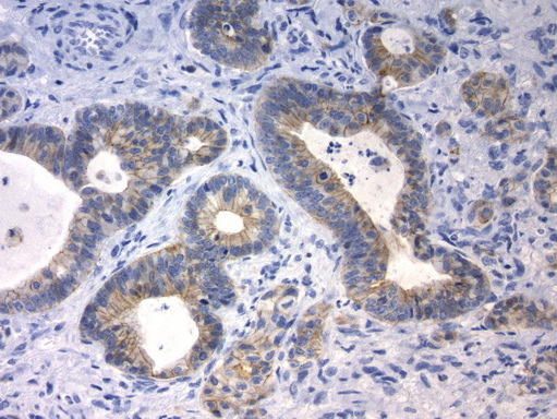
- Immunohistochemical staining of paraffin-embedded Carcinoma of Human prostate tissue using anti-CDH1 Mouse monoclonal antibody.(Heat-induced epitope retrieval by 1mM EDTA in 10mM Tris buffer (pH8.0) at 120C for 3 min, UM800076)(1:200)
- Validation comment
- IHC
- Submitted by
- OriGene (provider)
- Main image
- Experimental details
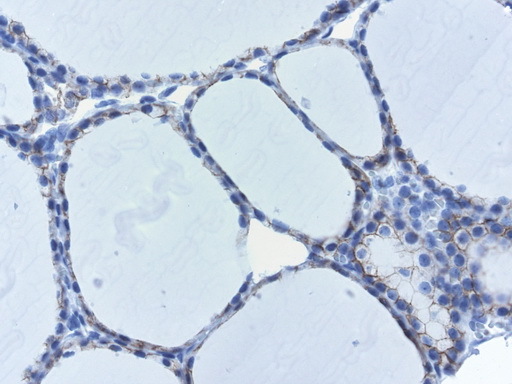
- Immunohistochemical staining of paraffin-embedded Carcinoma of Human thyroid tissue using anti-CDH1 mouse monoclonal antibody.(Heat-induced epitope retrieval by 1mM EDTA in 10mM Tris buffer (pH8.0) at 120C for 3 min, UM800076)(1:200)
- Validation comment
- IHC
- Submitted by
- OriGene (provider)
- Main image

- Experimental details
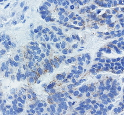
- Immunohistochemical staining of paraffin-embedded Adenocarcinoma of Human breast tissue using anti-CDH1 mouse monoclonal antibody. (Heat-induced epitope retrieval by 1mM EDTA in 10mM Tris buffer (pH8.0) at 120C for 3 min, UM800076)(1:200)
- Validation comment
- IHC
- Submitted by
- OriGene (provider)
- Main image

- Experimental details
- Immunohistochemical staining of paraffin-embedded Human prostate tissue using anti-CDH1 mouse monoclonal antibody. (Heat-induced epitope retrieval by 1mM EDTA in 10mM Tris buffer (pH8.0) at 120C for 3 min, UM800076)(1:200)
- Validation comment
- IHC
- Submitted by
- OriGene (provider)
- Main image

- Experimental details
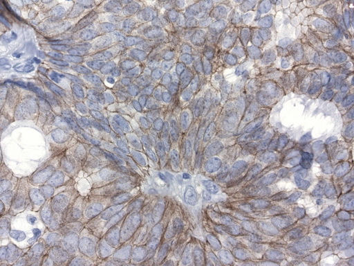
- Immunohistochemical staining of paraffin-embedded human thyroid using mouse anti-CDH1 clone UMAB184 (UM800076) at 1:200 with Polink2 Broad HRP DAB detection kit; pretreatment of tissue prior to stain with heat-induced epitope retrieval with GBI Accel pH 8.7 buffer using pressure chamber for 3 minutes at 110C is required for optimal staining. Shows membraneous staining.
- Validation comment
- IHC
- Submitted by
- OriGene (provider)
- Main image
- Experimental details
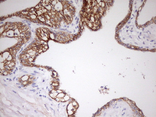
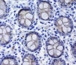
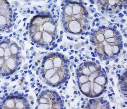
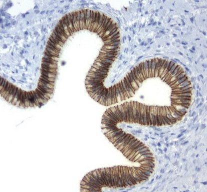
- Immunohistochemical staining of paraffin-embedded human small intestine using mouse anti-CDH1 clone UMAB184 (UM800076) at 1:200 with Polink2 Broad HRP DAB detection kit; pretreatment of tissue prior to stain with heat-induced epitope retrieval with GBI Accel pH 8.7 buffer using pressure chamber for 3 minutes at 110C is required for optimal staining. Shows mainly membranous staining and weak cytoplasmic staining.
- Validation comment
- IHC
- Submitted by
- OriGene (provider)
- Main image
- Experimental details
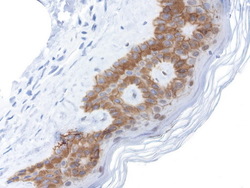
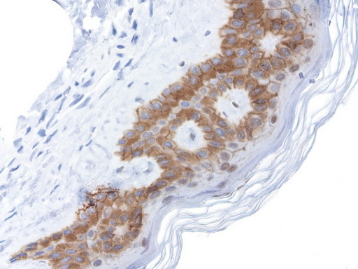
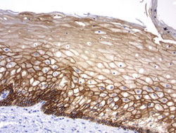
- Immunohistochemical staining of paraffin-embedded human skin using mouse anti-CDH1 clone UMAB184 (UM800076) at 1:200 with Polink2 Broad HRP DAB detection kit; pretreatment of tissue prior to stain with heat-induced epitope retrieval with GBI Accel pH 8.7 buffer using pressure chamber for 3 minutes at 110C is required for optimal staining. Shown here strong cytoplamic and membraneous staining on epithelial cells.
- Validation comment
- IHC
- Submitted by
- OriGene (provider)
- Main image
- Experimental details
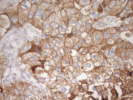
- Immunohistochemical staining of paraffin-embedded human prostate cancer using mouse anti-CDH1 clone UMAB184 (UM800076) at 1:200 with Polink2 Broad HRP DAB detection kit; pretreatment of tissue prior to stain with heat-induced epitope retrieval with GBI Accel pH 8.7 buffer using pressure chamber for 3 minutes at 110C is required for optimal staining. Shows membraneous staining of the tumor cells.
- Validation comment
- IHC

- Submitted by
- OriGene (provider)
- Main image
- Experimental details
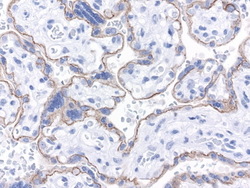
- Immunohistochemical staining of paraffin-embedded human placenta using mouse anti-CDH1 clone UMAB184 (UM800076) at 1:200 with Polink2 Broad HRP DAB detection kit; pretreatment of tissue prior to stain with heat-induced epitope retrieval with GBI Accel pH 8.7 buffer using pressure chamber for 3 minutes at 110C is required for optimal staining.
- Validation comment
- IHC
- Submitted by
- OriGene (provider)
- Main image
- Experimental details
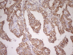
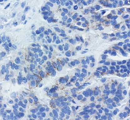
- Immunohistochemical staining of paraffin-embedded human ovarian cancer using mouse anti-CDH1 clone UMAB184 (UM800076) at 1:200 with Polink2 Broad HRP DAB detection kit; pretreatment of tissue prior to stain with heat-induced epitope retrieval with GBI Accel pH 8.7 buffer using pressure chamber for 3 minutes at 110C is required for optimal staining. Shows membraneous staining of the tumor cells.
- Validation comment
- IHC
- Submitted by
- OriGene (provider)
- Main image
- Experimental details
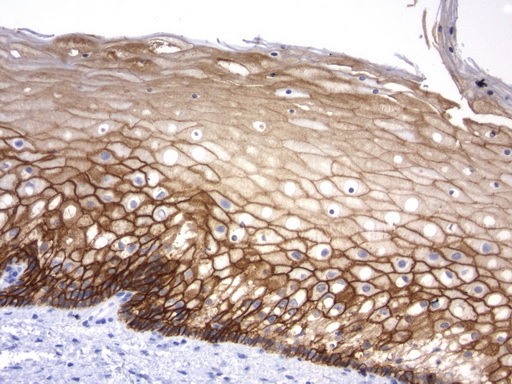
- Immunohistochemical staining of paraffin-embedded human normal cervix using mouse anti-CDH1 clone UMAB184 (UM800076) at 1:200 with Polink2 Broad HRP DAB detection kit; pretreatment of tissue prior to stain with heat-induced epitope retrieval with GBI Accel pH 8.7 buffer using pressure chamber for 3 minutes at 110C is required for optimal staining. Shown here strong cytoplamic and membraneous staining on squamous epithelial cells of the cervix.
- Validation comment
- IHC
- Submitted by
- OriGene (provider)
- Main image
- Experimental details
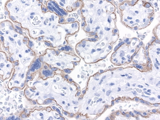
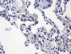
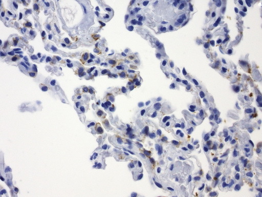
- Immunohistochemical staining of paraffin-embedded human lung using mouse anti-CDH1 clone UMAB184 (UM800076) at 1:200 with Polink2 Broad HRP DAB detection kit; pretreatment of tissue prior to stain with heat-induced epitope retrieval with GBI Accel pH 8.7 buffer using pressure chamber for 3 minutes at 110C is required for optimal staining. Stain shows membraneous staining.
- Validation comment
- IHC
- Submitted by
- OriGene (provider)
- Main image
- Experimental details
- Immunohistochemical staining of paraffin-embedded human endometrium using mouse anti-CDH1 clone UMAB184 (UM800076) at 1:200 with Polink2 Broad HRP DAB detection kit; pretreatment of tissue prior to stain with heat-induced epitope retrieval with GBI Accel pH 8.7 buffer using pressure chamber for 3 minutes at 110C is required for optimal staining. Stain shows membraneous staining on endometrium.
- Validation comment
- IHC
- Submitted by
- OriGene (provider)
- Main image

- Experimental details
- Immunohistochemical staining of paraffin-embedded human endocervical gland using mouse anti-CDH1 clone UMAB184 (UM800076) at 1:200 with Polink2 Broad HRP DAB detection kit; pretreatment of tissue prior to stain with heat-induced epitope retrieval with GBI Accel pH 8.7 buffer using pressure chamber for 3 minutes at 110C is required for optimal staining. Stain shows membraneous staining on epithelial cells of the gland.
- Validation comment
- IHC
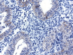
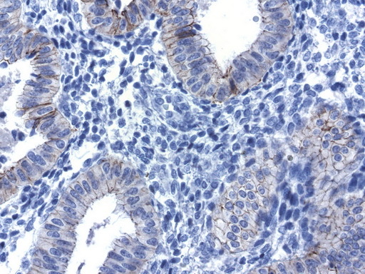
- Submitted by
- OriGene (provider)
- Main image

- Experimental details
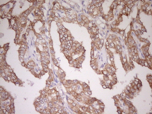
- Immunohistochemical staining of paraffin-embedded human colon cancer using mouse anti-CDH1 clone UMAB184 (UM800076) at 1:200 with Polink2 Broad HRP DAB detection kit; pretreatment of tissue prior to stain with heat-induced epitope retrieval with GBI Accel pH 8.7 buffer using pressure chamber for 3 minutes at 110C is required for optimal staining. Stain shows membraneous staining on tumor cells.
- Validation comment
- IHC
- Submitted by
- OriGene (provider)
- Main image

- Experimental details
- IHC staining of FFPE human breast cancer using anti-CDH1 clone UMAB184 mouse monoclonal antibody at 1:200 and detection with Polink2 Broad HRP DAB. UM800076 requires heat-induced epitope retrieval with Accel pH8.7. The image shows strong membranous and some weak cytoplasmic staining in the tumor cells.
- Validation comment
- IHC