Explore
 Validate
 Learn
Learn Western blot
 Flow cytometry
Antibody data
- Antibody Data
- Antigen structure
- References [2]
- Comments [0]
- Validations
- Flow cytometry [3]
Submit
Validation data
Reference
Comment
Report error
- Product number
- NBP1-42793 - Provider product page

- Provider
- Novus Biologicals
- Proper citation
- Novus Cat#NBP1-42793, RRID:AB_10005931
- Product name
- Mouse Monoclonal E-Cadherin Antibody
- Antibody type
- Monoclonal
- Description
- Protein A purified. The mouse monoclonal antibody 67A4 recognizes CD324/ E-cadherin, an approximately 100 kDa epithelial cell adhesion molecule, whose detection is important for determination of invasive potential of epithelial neoplasms. HLDA VIII
- Reactivity
- Human
- Host
- Mouse
- Isotype
- IgG
- Vial size
- 0.1 mg
- Concentration
- 1.0 mg/ml
- Storage
- Store at 4C. Do not freeze.
Submitted references Rapid resensitization of ASIC2a is conferred by three amino acid residues in the N terminus.
ASIC2a-dependent increase of ASIC3 surface expression enhances the sustained component of the currents.
Lee JS, Kweon HJ, Lee H, Suh BC
The Journal of general physiology 2019 Jul 1;151(7):944-953
The Journal of general physiology 2019 Jul 1;151(7):944-953
ASIC2a-dependent increase of ASIC3 surface expression enhances the sustained component of the currents.
Kweon HJ, Cho JH, Jang IS, Suh BC
BMB reports 2016 Oct;49(10):542-547
BMB reports 2016 Oct;49(10):542-547
No comments: Submit comment
Supportive validation
- Submitted by
- Novus Biologicals (provider)
- Main image

- Experimental details
- Flow Cytometry: E-Cadherin Antibody (67A4) [NBP1-42793] - Surface staining of HT-29 / SP2 cells with anti-CD324 (67A4) PE.

- Submitted by
- Novus Biologicals (provider)
- Main image

- Experimental details
- Flow Cytometry: E-Cadherin Antibody (67A4) [NBP1-42793] - Surface staining of HT-29 / SP2 cells with anti-CD324 (67A4) APC.

- Submitted by
- Novus Biologicals (provider)
- Main image

- Experimental details
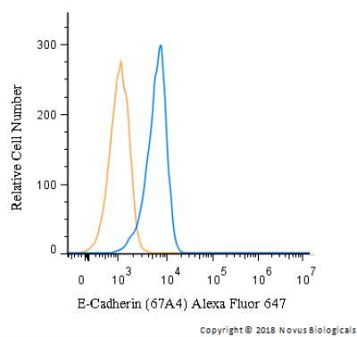
- Flow Cytometry: E-Cadherin Antibody (67A4) [NBP1-42793] - A surface stain was performed on A549 cells with E-Cadherin Antibody (67A4) NBP1-42793AF647 (blue) and a matched isotype control (orange). Cells were incubated in an antibody dilution of 5 ug/mL for 20 minutes at room temperature. Both antibodies were conjugated to Alexa Fluor 647.